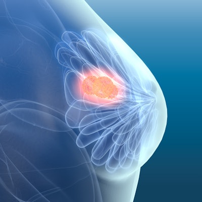
The American College of Radiology (ACR) has released updated breast cancer screening guidelines that underscore the need for early risk assessment -- especially among Black and Ashkenazi Jewish women.
The guidance calls for all women -- but these subgroups in particular -- to undergo a breast cancer risk workup by the age of 25. The recommendation was published May 3 in the Journal of the American College of Radiology. The guidance also reiterates that annual breast cancer screening should begin at age 40 for women at average risk of the disease.
Why the focus on Black women in particular and women of color in general getting early screening? The ACR listed the following reasons:
- Prior to age 50, women of color are 127% more likely to die of breast cancer; 72% more likely to be diagnosed with breast cancer; and 58% more likely to be diagnosed with advanced-stage breast cancer.
- Black women are 42% more likely to die from breast cancer despite equal incidence rates.
- Black women are less likely to be diagnosed with stage I breast cancer but twice as likely to die of early breast cancers.
- Black women have a two-fold higher risk of aggressive -- "triple-negative" -- breast tumors and a higher risk of BRCA1 and BRCA2 genetic mutations.
"The latest scientific evidence continues to point to earlier assessment as well as augmented and earlier-than-age-40 screening of many women -- particularly Black women and other minority women," Dr. Debra Monticciolo of Massachusetts General Hospital in Boston and co-founder and lead of the ACR's Screening Leadership Group, said in a statement released by the college. "These evidence-based updates should spur more-informed doctor-patient conversations and help providers save more lives."
The ACR also added the following updates to the guideline:
- Women with genetics-based increased risk (including BRCA1 carriers), those with a calculated lifetime risk of 20% or more, and those exposed to chest radiation at a young age are recommended to have MRI surveillance starting at ages 25 to 30. These women should start annual mammography at ages 25 to 40, depending on type of risk.
- Women diagnosed with breast cancer prior to age 50 or with a personal history of breast cancer and dense breasts should have annual supplemental breast MRI.
- High-risk women who desire supplemental screening -- but cannot undergo MRI screening -- should consider contrast-enhanced mammography.
National Cancer Institute Surveillance, Epidemiology, and End Results (SEER) data show that since mammography screening was implemented in the U.S. in the 1980s, the breast cancer death rate in women dropped 43%, the ACR noted.


![Examples of ultrasound findings and techniques. (A) Images in a 39-year-old male patient with a mass in the left thigh. The mass is heterogeneous on the B-mode US image (compared with the patient in D) and showed increased microvascularity (superb microvascular imaging [SMI]) and shear-wave elastography (SWE) values. Undifferentiated pleomorphic sarcoma was diagnosed at biopsy (with pleomorphic rhabdomyosarcoma in surgical specimen). (B) Images in an 18-year-old male patient with a mass in the left leg. The mass is hypoechoic on the B-mode image, with no other findings suggestive of malignancy. The lesion is in contact with the cortex of the tibia, which is slightly irregular. CT revealed a doubtful anteromedial tibial erosion. The microvascular study demonstrated high vascularization, suggestive of malignancy. Periosteal Ewing sarcoma was diagnosed with both histologic and immunohistochemical confirmation. (C) Images in a 69-year-old female patient with a lump growing on the outside of the left leg. Multiple SWE examinations were performed (please note the high values obtained in the measurements, whereas the color map highlights the stiffness relative to adjacent tissues). SMI showed areas of increased vascularization to target for sampling. Undifferentiated spindle cell sarcoma was diagnosed at biopsy, with residual leiomyosarcoma in the surgical specimen after neoadjuvant therapy. (D) Images in a 56-year-old female patient with a mass in the right thigh. The mass is heterogeneous at both B-mode ultrasound (similar to patient A) and MRI (coronal T2-weighted spectral attenuated inversion recovery [SPAIR]; T1-weighted pre-contrast and postcontrast imaging), which even shows uptake after the administration of paramagnetic contrast material, which is traditionally suggestive of malignancy. Low values at SMI and elastography are suggestive of benignity. Spindle cell lipoma was diagnosed at biopsy, with atypical spindle cell lipomatous tumor in the surgical specimen.](https://img.auntminnieeurope.com/mindful/smg/workspaces/default/uploads/2026/08/images-radiol250278fig2.APCFLSvX6p.jpg?auto=format%2Ccompress&fit=crop&h=100&q=70&w=100)


![Examples of ultrasound findings and techniques. (A) Images in a 39-year-old male patient with a mass in the left thigh. The mass is heterogeneous on the B-mode US image (compared with the patient in D) and showed increased microvascularity (superb microvascular imaging [SMI]) and shear-wave elastography (SWE) values. Undifferentiated pleomorphic sarcoma was diagnosed at biopsy (with pleomorphic rhabdomyosarcoma in surgical specimen). (B) Images in an 18-year-old male patient with a mass in the left leg. The mass is hypoechoic on the B-mode image, with no other findings suggestive of malignancy. The lesion is in contact with the cortex of the tibia, which is slightly irregular. CT revealed a doubtful anteromedial tibial erosion. The microvascular study demonstrated high vascularization, suggestive of malignancy. Periosteal Ewing sarcoma was diagnosed with both histologic and immunohistochemical confirmation. (C) Images in a 69-year-old female patient with a lump growing on the outside of the left leg. Multiple SWE examinations were performed (please note the high values obtained in the measurements, whereas the color map highlights the stiffness relative to adjacent tissues). SMI showed areas of increased vascularization to target for sampling. Undifferentiated spindle cell sarcoma was diagnosed at biopsy, with residual leiomyosarcoma in the surgical specimen after neoadjuvant therapy. (D) Images in a 56-year-old female patient with a mass in the right thigh. The mass is heterogeneous at both B-mode ultrasound (similar to patient A) and MRI (coronal T2-weighted spectral attenuated inversion recovery [SPAIR]; T1-weighted pre-contrast and postcontrast imaging), which even shows uptake after the administration of paramagnetic contrast material, which is traditionally suggestive of malignancy. Low values at SMI and elastography are suggestive of benignity. Spindle cell lipoma was diagnosed at biopsy, with atypical spindle cell lipomatous tumor in the surgical specimen.](https://img.auntminnieeurope.com/mindful/smg/workspaces/default/uploads/2026/08/images-radiol250278fig2.APCFLSvX6p.jpg?auto=format%2Ccompress&fit=crop&h=112&q=70&w=112)