Microwave ablation can effectively treat papillary thyroid microcarcinoma using ultrasound to identify capsular invasion -- an approach which has been typically avoided due to safety concerns, suggest findings published on 7 March in Radiology.
A team led by Lin Zheng from the People's Liberation Army General Hospital in Beijing, China, found that treating thyroid microcarcinoma with microwave ablation and ultrasound showed comparable short-term efficacy whether or not the disease included capsular invasion (that is, when the tumor extends through the entire thyroid capsule) -- a result that counters the traditional assumption that using ultrasound in this way is not a safe for the procedure, the group noted.
"Our results indicated that the treatment outcomes of microwave ablation on papillary thyroid microcarcinoma both with and without capsular invasion were similar," the investigators wrote.
Most detected papillary thyroid microcarcinomas are of low risk and have a favorable prognosis, according to the authors. But current guidelines recommend surgical resection or active surveillance for these cases. In contrast, microwave ablation is a minimally invasive method that has shown promise in previous studies for treating such carcinomas -- although Zheng and colleagues noted that using it to treat papillary thyroid microcarcinomas with aggressive features remains under debate.
Capsular invasion has been previously identified as an independent risk factor for thyroid lymph node metastasis. Yet papillary thyroid microcarcinomas with ultrasound-detected capsular invasion have been excluded from thermal ablation treatment due to concerns about increasing complications post treatment.
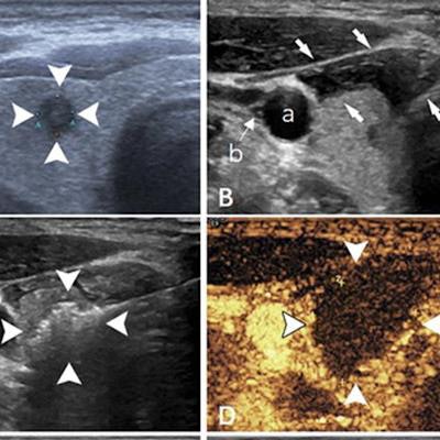
 Ultrasound images show papillary thyroid carcinoma in the right lobe of the thyroid in a 36-year-old man. (A) Imaging shows the tumor pre-ablation (arrowheads). (B) The hydrodissection technique (arrows) was used to protect the carotid artery (a) and vagus nerve (b). (C) Imaging shows a hyperechoic pattern in the tumor during the ablation procedure (arrowheads). (D) Postablation contrast-enhanced imaging shows no enhancement in the tumor area (arrowheads). (E) Imaging shows the ablation zone six months after ablation (arrowheads). (F) The ablation area has almost disappeared one year after ablation. Images and caption courtesy of Radiology.
Zheng and co-authors sought to compare the feasibility, effectiveness, and safety of microwave ablation for treating these microcarcinomas with and without ultrasound-detected capsular invasion via a study that included data from 460 patients. Of these, 82 underwent microwave ablation with ultrasound-guided detection of capsular invasion while the remaining 378 did not. The capsular invasion group had follow-up at 20 months while the noninvasion group had follow-up at 21 months.
The team found comparable success rates for microwave ablation with ultrasound for both groups, at 99% for the capsular invasion group and 100% for the non-invasion group. It also reported only one complication (1%) in the capsular invasion group compared with 11 in the noninvasion group (3%). On follow-up, the team reported clear shrinkage of the ablation zone in both groups, at 97% in the capsular invasion group and 96% in the noninvasion group.
All participants with complications recovered within three months during the follow-up period, and none developed permanent voice hoarseness, which can be a complication of thyroid cancer treatment, according to Zheng and colleagues.
The authors recommended further studies with more participants and longer follow-up to validate their findings, and they added that further comparison with active surveillance would be of particular importance to clarify the clinical impact of ablation therapy with ultrasound.