There are particular CT findings that correlate with histologic data and identify early interstitial lung abnormalities, but sometimes findings that appear normal on CT are abnormal on histopathology, a Belgian-led research team has found.
The results highlight the fact that more work needs to be done to effectively assess lung abnormalities that could indicate interstitial lung disease, according to a study published on 20 December in Radiology.
"Future studies are required to further refine and validate the concept of interstitial lung abnormalities and their relation to the genesis and progression of interstitial lung disease," wrote Stijn Verleden, PhD, a post-doctoral researcher at Antwerp University and Katholieke Universiteit Leuven (KU Leuven) in Belgium, and colleagues.
Although interstitial lung abnormalities found on lung CT scans can indicate early interstitial lung disease, the condition's histopathologic correlates to imaging results remain unclear, they noted. To address the knowledge gap, they conducted a study that included data from eight lung specimens taken between 2010 and 2021 from six donors with radiologically confirmed interstitial lung disease. The tissue was inflated, frozen, and scanned with CT and micro-CT imaging, then sampled via core biopsy. The group evaluated any associations between the imaging and histopathologic findings.
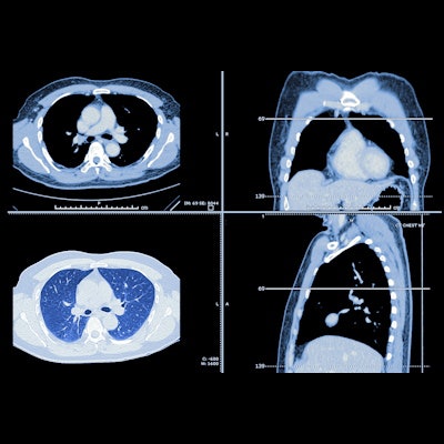
CT findings included ground-glass opacification, reticulation, and bronchiectasis, while micro-CT and histopathologic examination showed that lung abnormalities were often paraseptal and associated with fibrosis and lymphocytic inflammation.
Of concern, however, is that histopathologic results showed degrees of fibrosis that appeared normal on CT imaging.
"[We] have demonstrated that the degree of fibrosis in relatively normal-appearing lung regions with histopathologic evidence of moderate fibrosis ... and inflammation ... was underestimated at CT," the authors wrote.
The study findings underscore how it's "difficult to effectively characterize interstitial lung abnormalities with current imaging techniques," wrote Dr. Jean Jeudy of the University of Maryland in Baltimore in an accompanying commentary, cautioning that, when it comes to interstitial lung abnormalities, it could be that "the more we learn, the less we know."
"Patients with advanced features of fibrosis have been shown to be at highest risk for progression and adverse outcomes," he noted. "However, even patients with mild disease may have underlying fibrosis that could be missed with CT."
Yet it's likely that further imaging research will improve the diagnosis of early interstitial lung disease, according to Jeudy.
"Newer advances in scanner technology and artificial intelligence will improve the accuracy of our imaging," he concluded. "With improved imaging biomarkers, the definitive goal will be to identify which patients will develop progressive disease and why."


![Examples of ultrasound findings and techniques. (A) Images in a 39-year-old male patient with a mass in the left thigh. The mass is heterogeneous on the B-mode US image (compared with the patient in D) and showed increased microvascularity (superb microvascular imaging [SMI]) and shear-wave elastography (SWE) values. Undifferentiated pleomorphic sarcoma was diagnosed at biopsy (with pleomorphic rhabdomyosarcoma in surgical specimen). (B) Images in an 18-year-old male patient with a mass in the left leg. The mass is hypoechoic on the B-mode image, with no other findings suggestive of malignancy. The lesion is in contact with the cortex of the tibia, which is slightly irregular. CT revealed a doubtful anteromedial tibial erosion. The microvascular study demonstrated high vascularization, suggestive of malignancy. Periosteal Ewing sarcoma was diagnosed with both histologic and immunohistochemical confirmation. (C) Images in a 69-year-old female patient with a lump growing on the outside of the left leg. Multiple SWE examinations were performed (please note the high values obtained in the measurements, whereas the color map highlights the stiffness relative to adjacent tissues). SMI showed areas of increased vascularization to target for sampling. Undifferentiated spindle cell sarcoma was diagnosed at biopsy, with residual leiomyosarcoma in the surgical specimen after neoadjuvant therapy. (D) Images in a 56-year-old female patient with a mass in the right thigh. The mass is heterogeneous at both B-mode ultrasound (similar to patient A) and MRI (coronal T2-weighted spectral attenuated inversion recovery [SPAIR]; T1-weighted pre-contrast and postcontrast imaging), which even shows uptake after the administration of paramagnetic contrast material, which is traditionally suggestive of malignancy. Low values at SMI and elastography are suggestive of benignity. Spindle cell lipoma was diagnosed at biopsy, with atypical spindle cell lipomatous tumor in the surgical specimen.](https://img.auntminnieeurope.com/mindful/smg/workspaces/default/uploads/2026/08/images-radiol250278fig2.APCFLSvX6p.jpg?auto=format%2Ccompress&fit=crop&h=100&q=70&w=100)


![Examples of ultrasound findings and techniques. (A) Images in a 39-year-old male patient with a mass in the left thigh. The mass is heterogeneous on the B-mode US image (compared with the patient in D) and showed increased microvascularity (superb microvascular imaging [SMI]) and shear-wave elastography (SWE) values. Undifferentiated pleomorphic sarcoma was diagnosed at biopsy (with pleomorphic rhabdomyosarcoma in surgical specimen). (B) Images in an 18-year-old male patient with a mass in the left leg. The mass is hypoechoic on the B-mode image, with no other findings suggestive of malignancy. The lesion is in contact with the cortex of the tibia, which is slightly irregular. CT revealed a doubtful anteromedial tibial erosion. The microvascular study demonstrated high vascularization, suggestive of malignancy. Periosteal Ewing sarcoma was diagnosed with both histologic and immunohistochemical confirmation. (C) Images in a 69-year-old female patient with a lump growing on the outside of the left leg. Multiple SWE examinations were performed (please note the high values obtained in the measurements, whereas the color map highlights the stiffness relative to adjacent tissues). SMI showed areas of increased vascularization to target for sampling. Undifferentiated spindle cell sarcoma was diagnosed at biopsy, with residual leiomyosarcoma in the surgical specimen after neoadjuvant therapy. (D) Images in a 56-year-old female patient with a mass in the right thigh. The mass is heterogeneous at both B-mode ultrasound (similar to patient A) and MRI (coronal T2-weighted spectral attenuated inversion recovery [SPAIR]; T1-weighted pre-contrast and postcontrast imaging), which even shows uptake after the administration of paramagnetic contrast material, which is traditionally suggestive of malignancy. Low values at SMI and elastography are suggestive of benignity. Spindle cell lipoma was diagnosed at biopsy, with atypical spindle cell lipomatous tumor in the surgical specimen.](https://img.auntminnieeurope.com/mindful/smg/workspaces/default/uploads/2026/08/images-radiol250278fig2.APCFLSvX6p.jpg?auto=format%2Ccompress&fit=crop&h=112&q=70&w=112)