Transthoracic echocardiography is helpful in determining mortality risk for patients with COVID-19, according to research published on 21 July in the European Journal of Clinical Investigation.
Researchers led by Dr. Angelo Silverio from the University of Salerno in Italy found that transthoracic echo calculations of left ventricular ejection fraction and tricuspid annular plane systolic excursion are independently associated with in-hospital mortality for COVID-19 patients.
They also found a significantly higher risk of mortality in patients with acute respiratory distress syndrome (ARDS) compared with those without ARDS.
"Cardiovascular complications can negatively impact on outcomes of patients with COVID-19," the study authors wrote. "Clinical and echocardiographic parameters might help to identify patients at higher risk for in-hospital mortality."
Italy's number of active COVID-19 cases was one of the highest in the world during the early months of the COVID-19 pandemic. As of July 20, the country has had nearly 4.3 million total cases, including 127,874 deaths.
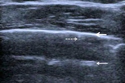
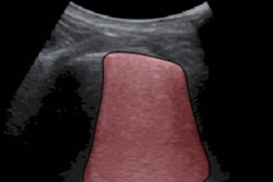
Previous research has touted the wide availability and feasibility of transthoracic echocardiography (TTE) in diagnosing patients in critical care settings with suspected or confirmed cardiac disease. However, using TTE for COVID-19 can be challenging, since it requires close contact with patients. Because of this, echocardiographers may be at risk for infection.
The current recommendation for using TTE for COVID patients is to use it only on a case-by-case basis and if it is deemed essential for patient care.
Researchers said that the role of TTE in classifying risk of COVID-19 patients has been "poorly" investigated. They wanted to look at clinical characteristics of hospitalized patients and find out the association between these characteristics and echocardiographic parameters with in-hospital mortality.
The team looked at 1,401 COVID-19 patients from seven Italian medical centers from March to April 2020, 226 of whom underwent TTE within 48 hours of being admitted. Each referral for TTE was confirmed by a cardiologist to minimize exposure of COVID-19 to staff. In-hospital death occurred in 68 cases. The rest of the patients were discharged after recovering.
The researchers found that reduced left ventricular ejection fraction, reduced tricuspid annular plane systolic excursion, and ARDS were independently associated with in-hospital mortality (p < 0.001).
| Risk probability for in-hospital mortality of COVID-19 patients measured with TTE | |
| Clinical characteristics | Hazard ratio |
| Patients with ARDS | 7.66 |
| Tricuspid annular plane systolic excursion ≤ 17 mm | 5.08 |
| Left ventricular ejection fraction ≤ 50% | 4.06 |
Males had a higher prevalence of death than females in the study. Out of the 141 male patients included, 51 died in their respective hospital settings (p = 0.016).
In patients with reduced left ventricular ejection fraction, COVID-19 could act like a precipitating factor and rapidly deteriorate the patients' clinical status, the researchers noted.
"In fact, previous studies demonstrated the association of concomitant cardiac disease with outcome in patients infected by SARS-CoV-2," the authors wrote.
The team said their preliminary findings need to be confirmed in larger, prospective studies.