Dear AuntMinnieEurope Member,
Our editorial team always bears in mind that clinical images are the lifeblood of readers, so I'm delighted to report that this week's top two articles contain some stunning images and cases.
First up, French experts in ob/gyn imaging have shared their experiences of endometriosis. They think it's particularly important for radiologists to know about the acute complications of endometriomas and to look out for characteristic T1-weighted fat-suppressed foci. Find out more in the MRI Community.
Second, we feature new work from Latin America, where the use of silicone for cosmetic purposes is growing fast. Imaging can warn surgeons about the level of tissue compromise, show the affected planes and the amount of biopolymers injected, and demonstrate their areas in the case of material migration. It's vital for radiologists to learn to recognize the images that they generate, Colombian specialists say. Go to the Women's Imaging Community for the full story.
Radiology Across Borders (RAB) is a very worthwhile charity that organizes hands-on training, particularly in developing nations. We published a feature about them in March 2017, and now we've posted an update based on an interview with Dr. Suresh de Silva, co-founder of RAB and an oncological/body imaging consultant. It's heartening to see that de Silva and his colleagues are still going strong in the pandemic.
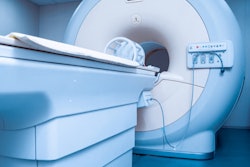
Asymmetric Parkinson's disease can be very distressing to patients because the symptoms are much more severe on one side of the body. A Spanish-led team has found that focused ultrasound is showing considerable promise in this area.
In another ultrasound article, researchers from Tours, France, have developed a thyroid cancer screening technique that hit all the right notes. Their method can noninvasively detect abnormally stiff tissues when participants sing the "eeee" sound. Don't miss our uplifting report.