
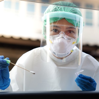
VIDI, the leading private radiology group in France, has stated that it will ensure the availability of personal protective equipment (PPE) across its network of centers to protect patients from infection with the novel coronavirus.
The group is responding to recent concerns raised by Health Minister Olivier Veran, who stated that patients who may have serious diseases such as cancer are avoiding going to centers for diagnosis or treatment due to fears about the virus. Such delays could potentially affect patient outcomes, VIDI noted in a press release issued on 27 April.
VIDI said it has been quick to ensure the availability of protective gear so that patients can be examined in the safest conditions while staff are also protected.
The reception of patients at VIDI centers has been redesigned to shorten waiting times and respect social distancing, while time between exams allows for disinfection of imaging equipment, according to the group.
VIDI centers are also contacting patients who had to cancel exams due to the lockdown to remind them to reschedule imaging, while reassuring them of safety measures. Last week, 96% of patients turned up for their appointments, the group noted. It stated that its radiologists were available to respond to the health minister's call to patients to "get themselves treated."


![Overview of the study design. (A) The fully automated deep learning framework was developed to estimate body composition (BC) (defined as subcutaneous adipose tissue [SAT] in liters; visceral adipose tissue [VAT] in liters; skeletal muscle [SM] in liters; SM fat fraction [SMFF] as a percentage; and intramuscular adipose tissue [IMAT] in deciliters) from MRI. The fully automated framework comprised one model (model 1) to quantify different BC measures (SAT, VAT, SM, SMFF, and IMAT) as three-dimensional (3D) measures from whole-body MRI scans. The second model (model 2) was trained to identify standardized anatomic landmarks along the craniocaudal body axis (z coordinate field), which allowed for subdividing the whole-body measures into different subregions typically examined on clinical routine MRI scans (chest, abdomen, and pelvis). (B) BC was quantified from whole-body MRI in over 66,000 individuals from two large population-based cohort studies, the UK Biobank (UKB) (36,317 individuals) and the German National Cohort (NAKO) (30,291 individuals). Bar graphs show age distribution by sex and cohort. BMI = body mass index. (C) After the performance assessment of the fully automated framework, the change in BC measures, distributions, and profiles across age decades were investigated. Age-, sex-, and height-adjusted body composition reference curves were calculated and made publicly available in a web-based z-score calculator (https://circ-ml.github.io).](https://img.auntminnieeurope.com/mindful/smg/workspaces/default/uploads/2026/05/body-comp.XgAjTfPj1W.jpg?auto=format%2Ccompress&fit=crop&h=112&q=70&w=112)


