Shielding may be on the way out in x-ray, but it's in vogue in ultrasound. A Dutch radiologist has worked with one of the big four imaging manufacturers and technical experts to develop a screen designed to protect staff and patients from COVID-19.
Dr. Herma Holscher, a pediatric radiologist at Haga Teaching Hospital / Juliana Children's Hospital in The Hague, had the initial idea, and the screen is now being used in her department.
"Super happy with our prototype screen," says Dr. Herma Holscher."The prototype screen aims to make the ultrasound environment safer, especially for children, their parents, and the radiologist. This is precisely because today's nonemergency can be tomorrow's emergency care," she noted in a LinkedIn posting.
She developed the screen with Siemens Healthineers. It was custom-built for the hospital by a technical company called Van Vliet Medical in Almere, the Netherlands.
An artist's impression of how the screen works. Copyright: Siemens Healthineers."It took us one week to have it all working. It is a prototype made from plexiglass with two holes (just like an incubator wall, for small interventions), and it is on wheels. So it really is as simple as that!" Holscher told AuntMinnieEurope.com.
The ultrasound screen is available to purchase from Van Vliet Medical at a cost of 1,396 euros, excluding tax. The product is not patented. "I feel, and this feeling is supported by Siemens and Van Vliet Medical, that we should not make a profit out of this crisis; any good idea should be shared. The price of the product is the cost price," she said.
Haga Teaching hospital has nearly 4,000 staff and around 250 medical specialists. It is one of 19 large teaching hospitals in the Netherlands.
Along with Siemens, Holscher has also developed a dedicated thoracic x-ray device for children.
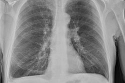
The dedicated thoracic x-ray scanner is useful for children who are too young to do breath-hold. Images courtesy of Dr. Herma Holscher.
"With a mirror, technicians can see the breathing of the child to make proper inspiration -- and on occasion, expiration -- x-rays in children who are too young to do breath-hold," she explained.

![Examples of ultrasound findings and techniques. (A) Images in a 39-year-old male patient with a mass in the left thigh. The mass is heterogeneous on the B-mode US image (compared with the patient in D) and showed increased microvascularity (superb microvascular imaging [SMI]) and shear-wave elastography (SWE) values. Undifferentiated pleomorphic sarcoma was diagnosed at biopsy (with pleomorphic rhabdomyosarcoma in surgical specimen). (B) Images in an 18-year-old male patient with a mass in the left leg. The mass is hypoechoic on the B-mode image, with no other findings suggestive of malignancy. The lesion is in contact with the cortex of the tibia, which is slightly irregular. CT revealed a doubtful anteromedial tibial erosion. The microvascular study demonstrated high vascularization, suggestive of malignancy. Periosteal Ewing sarcoma was diagnosed with both histologic and immunohistochemical confirmation. (C) Images in a 69-year-old female patient with a lump growing on the outside of the left leg. Multiple SWE examinations were performed (please note the high values obtained in the measurements, whereas the color map highlights the stiffness relative to adjacent tissues). SMI showed areas of increased vascularization to target for sampling. Undifferentiated spindle cell sarcoma was diagnosed at biopsy, with residual leiomyosarcoma in the surgical specimen after neoadjuvant therapy. (D) Images in a 56-year-old female patient with a mass in the right thigh. The mass is heterogeneous at both B-mode ultrasound (similar to patient A) and MRI (coronal T2-weighted spectral attenuated inversion recovery [SPAIR]; T1-weighted pre-contrast and postcontrast imaging), which even shows uptake after the administration of paramagnetic contrast material, which is traditionally suggestive of malignancy. Low values at SMI and elastography are suggestive of benignity. Spindle cell lipoma was diagnosed at biopsy, with atypical spindle cell lipomatous tumor in the surgical specimen.](https://img.auntminnieeurope.com/mindful/smg/workspaces/default/uploads/2026/08/images-radiol250278fig2.APCFLSvX6p.jpg?auto=format%2Ccompress&fit=crop&h=100&q=70&w=100)


![Examples of ultrasound findings and techniques. (A) Images in a 39-year-old male patient with a mass in the left thigh. The mass is heterogeneous on the B-mode US image (compared with the patient in D) and showed increased microvascularity (superb microvascular imaging [SMI]) and shear-wave elastography (SWE) values. Undifferentiated pleomorphic sarcoma was diagnosed at biopsy (with pleomorphic rhabdomyosarcoma in surgical specimen). (B) Images in an 18-year-old male patient with a mass in the left leg. The mass is hypoechoic on the B-mode image, with no other findings suggestive of malignancy. The lesion is in contact with the cortex of the tibia, which is slightly irregular. CT revealed a doubtful anteromedial tibial erosion. The microvascular study demonstrated high vascularization, suggestive of malignancy. Periosteal Ewing sarcoma was diagnosed with both histologic and immunohistochemical confirmation. (C) Images in a 69-year-old female patient with a lump growing on the outside of the left leg. Multiple SWE examinations were performed (please note the high values obtained in the measurements, whereas the color map highlights the stiffness relative to adjacent tissues). SMI showed areas of increased vascularization to target for sampling. Undifferentiated spindle cell sarcoma was diagnosed at biopsy, with residual leiomyosarcoma in the surgical specimen after neoadjuvant therapy. (D) Images in a 56-year-old female patient with a mass in the right thigh. The mass is heterogeneous at both B-mode ultrasound (similar to patient A) and MRI (coronal T2-weighted spectral attenuated inversion recovery [SPAIR]; T1-weighted pre-contrast and postcontrast imaging), which even shows uptake after the administration of paramagnetic contrast material, which is traditionally suggestive of malignancy. Low values at SMI and elastography are suggestive of benignity. Spindle cell lipoma was diagnosed at biopsy, with atypical spindle cell lipomatous tumor in the surgical specimen.](https://img.auntminnieeurope.com/mindful/smg/workspaces/default/uploads/2026/08/images-radiol250278fig2.APCFLSvX6p.jpg?auto=format%2Ccompress&fit=crop&h=112&q=70&w=112)