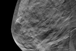
The performance of full-field digital mammography (FFDM) compared with film-screen mammography for microcalcifications was something of a mixed bag in a new study. FFDM had better detection of high-grade ductal carcinoma in situ (DCIS) but also a higher recall rate, according to research published online on 28 January in Radiology.
The new study looked at two decades of mammography screening results from hundreds of thousands of women in the Netherlands who participated in the country's breast screening program, which switched from film to full-field digital mammography between 2009 and 2010. The findings demonstrate that newer imaging technologies can have a big impact on early breast cancer detection.
"More than half of DCIS related to suspicious microcalcifications in our study showed high-grade histopathologic characteristics," wrote the authors, led by Dr. Jacky Luiten from the Elisabeth-Tweesteden Hospital in the Netherlands. "As the detection and subsequent treatment of high-grade DCIS may reduce further development of high-grade invasive carcinoma, histologic analysis of screening-detected microcalcifications carries substantial clinical value."
The researchers used data from more than 810,000 women who participated in the Dutch biennial mammography screening program between 1997 and 2017. The number of women with breast screening increased from 48,721 in the first studied year to 131,757 in the last.
The switch to full-field digital mammography between 2009 and 2010 resulted in the detection of more high-risk DCIS lesions and more cancer diagnoses. Detection of DCIS increased from 0.9 identified cancers per 1,000 screening examinations in the 2D film era to 1.7 identified cancers in the digital era. The rise in DCIS detection was significant in multiple statistical analyses.
But recalls for suspicious microcalcifications rose substantially after the switch to full-field digital mammography. Clinicians recalled 51 women for suspicious microcalcifications in the first studied screening year but 680 women in the last one.
| Analog vs. digital mammography in the Netherlands breast screening program | ||
| Film-screen mammography | FFDM | |
| Detection of DCIS per 1,000 exams | 0.9 lesions | 1.7 lesions |
| Recall rate | 0.1% | 0.5% |
| DCIS recalls per 1,000 exams | 0.3 recalls | 1.1 recalls |
| Positive predictive value for recall of microcalcifications | 51% | 33% |
The researchers hypothesized the rise in recall rates and identification of DCIS could signal that full-field digital mammography is better than film for finding suspicious microcalcifications. However, it could also result in more women getting tested and treated for low-risk lesions.
"Characterizing radiologic features of suspicious microcalcifications to search for patterns that correlate with more aggressive underlying disease might be a desirable next step toward a more selective use of [stereotactic core needle biopsy] in case of screening-detected calcifications," the authors wrote. "Because it is not yet clear to what extent histologic features of DCIS can be estimated by the patterns of microcalcifications alone, [biopsy] is still considered mandatory in the workup of these lesions to date."


![Examples of ultrasound findings and techniques. (A) Images in a 39-year-old male patient with a mass in the left thigh. The mass is heterogeneous on the B-mode US image (compared with the patient in D) and showed increased microvascularity (superb microvascular imaging [SMI]) and shear-wave elastography (SWE) values. Undifferentiated pleomorphic sarcoma was diagnosed at biopsy (with pleomorphic rhabdomyosarcoma in surgical specimen). (B) Images in an 18-year-old male patient with a mass in the left leg. The mass is hypoechoic on the B-mode image, with no other findings suggestive of malignancy. The lesion is in contact with the cortex of the tibia, which is slightly irregular. CT revealed a doubtful anteromedial tibial erosion. The microvascular study demonstrated high vascularization, suggestive of malignancy. Periosteal Ewing sarcoma was diagnosed with both histologic and immunohistochemical confirmation. (C) Images in a 69-year-old female patient with a lump growing on the outside of the left leg. Multiple SWE examinations were performed (please note the high values obtained in the measurements, whereas the color map highlights the stiffness relative to adjacent tissues). SMI showed areas of increased vascularization to target for sampling. Undifferentiated spindle cell sarcoma was diagnosed at biopsy, with residual leiomyosarcoma in the surgical specimen after neoadjuvant therapy. (D) Images in a 56-year-old female patient with a mass in the right thigh. The mass is heterogeneous at both B-mode ultrasound (similar to patient A) and MRI (coronal T2-weighted spectral attenuated inversion recovery [SPAIR]; T1-weighted pre-contrast and postcontrast imaging), which even shows uptake after the administration of paramagnetic contrast material, which is traditionally suggestive of malignancy. Low values at SMI and elastography are suggestive of benignity. Spindle cell lipoma was diagnosed at biopsy, with atypical spindle cell lipomatous tumor in the surgical specimen.](https://img.auntminnieeurope.com/mindful/smg/workspaces/default/uploads/2026/08/images-radiol250278fig2.APCFLSvX6p.jpg?auto=format%2Ccompress&fit=crop&h=100&q=70&w=100)


![Examples of ultrasound findings and techniques. (A) Images in a 39-year-old male patient with a mass in the left thigh. The mass is heterogeneous on the B-mode US image (compared with the patient in D) and showed increased microvascularity (superb microvascular imaging [SMI]) and shear-wave elastography (SWE) values. Undifferentiated pleomorphic sarcoma was diagnosed at biopsy (with pleomorphic rhabdomyosarcoma in surgical specimen). (B) Images in an 18-year-old male patient with a mass in the left leg. The mass is hypoechoic on the B-mode image, with no other findings suggestive of malignancy. The lesion is in contact with the cortex of the tibia, which is slightly irregular. CT revealed a doubtful anteromedial tibial erosion. The microvascular study demonstrated high vascularization, suggestive of malignancy. Periosteal Ewing sarcoma was diagnosed with both histologic and immunohistochemical confirmation. (C) Images in a 69-year-old female patient with a lump growing on the outside of the left leg. Multiple SWE examinations were performed (please note the high values obtained in the measurements, whereas the color map highlights the stiffness relative to adjacent tissues). SMI showed areas of increased vascularization to target for sampling. Undifferentiated spindle cell sarcoma was diagnosed at biopsy, with residual leiomyosarcoma in the surgical specimen after neoadjuvant therapy. (D) Images in a 56-year-old female patient with a mass in the right thigh. The mass is heterogeneous at both B-mode ultrasound (similar to patient A) and MRI (coronal T2-weighted spectral attenuated inversion recovery [SPAIR]; T1-weighted pre-contrast and postcontrast imaging), which even shows uptake after the administration of paramagnetic contrast material, which is traditionally suggestive of malignancy. Low values at SMI and elastography are suggestive of benignity. Spindle cell lipoma was diagnosed at biopsy, with atypical spindle cell lipomatous tumor in the surgical specimen.](https://img.auntminnieeurope.com/mindful/smg/workspaces/default/uploads/2026/08/images-radiol250278fig2.APCFLSvX6p.jpg?auto=format%2Ccompress&fit=crop&h=112&q=70&w=112)