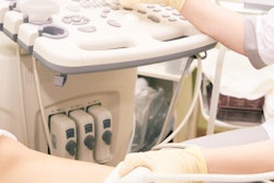
An astronaut on the International Space Station using an onboard ultrasound scanner to research the effects of space flight instead detected venous thrombosis in the individual's own body, according to an article published in the 2 January issue of the New England Journal of Medicine.
Two months into a six-month mission on the International Space Station, the astronaut identified a potential blood clot while performing a self-directed ultrasound exam as part of a vascular research study. A follow-up ultrasound exam performed by the astronaut with guidance from two radiologists on Earth confirmed that the blood clot was a deep vein thrombosis (DVT) in the jugular vein of the neck (NEJM, 2 January 2020, Vol. 382:1, pp. 89-90).
The International Space Station. Image courtesy of NASA.Treatment of the condition typically entails taking blood-thinning medication for several months to prevent the clot from growing and moving to a different part of the body, noted co-author Dr. Stephen Moll in a statement released by the University of North Carolina School of Medicine. Moll is a professor of medicine at the university and a member of its Blood Research Center.
"There is some risk when taking blood thinners that if an injury occurs, it could cause internal bleeding that is difficult to stop. ... Knowing there are no emergency rooms in space, we had to weigh our options very carefully," he said.
Moll discussed treatment options with a team of NASA physicians and finally advised the astronaut to take a blood thinner for approximately 90 days, with regular ultrasound exams guided by radiologists to monitor the blood clot. The medical team also advised the astronaut to stop taking the blood thinner four days before the journey back home to Earth due to the high physical demands involved in the reentry process.
After landing on Earth, the astronaut underwent a point-of-care ultrasound exam that indicated further treatment for DVT was not required. The astronaut remained asymptomatic on a follow-up exam six months after returning to Earth, the authors found.
The case of DVT in spaceflight underscores the complexities of space medicine, which, for this astronaut, included patient-performed, radiologist-guided ultrasonography, Moll and colleagues noted. The case revealed gaps in current understanding of circulatory and hemostatic physiology in space, as well as the need for more research on how blood and blood clots behave in space.
"How do you minimize risk for DVT?" Moll said. "Should there be more medications for it kept on the International Space Station? All of these questions need answering, especially with the plan that astronauts will embark on longer missions to the moon and Mars."


![Examples of ultrasound findings and techniques. (A) Images in a 39-year-old male patient with a mass in the left thigh. The mass is heterogeneous on the B-mode US image (compared with the patient in D) and showed increased microvascularity (superb microvascular imaging [SMI]) and shear-wave elastography (SWE) values. Undifferentiated pleomorphic sarcoma was diagnosed at biopsy (with pleomorphic rhabdomyosarcoma in surgical specimen). (B) Images in an 18-year-old male patient with a mass in the left leg. The mass is hypoechoic on the B-mode image, with no other findings suggestive of malignancy. The lesion is in contact with the cortex of the tibia, which is slightly irregular. CT revealed a doubtful anteromedial tibial erosion. The microvascular study demonstrated high vascularization, suggestive of malignancy. Periosteal Ewing sarcoma was diagnosed with both histologic and immunohistochemical confirmation. (C) Images in a 69-year-old female patient with a lump growing on the outside of the left leg. Multiple SWE examinations were performed (please note the high values obtained in the measurements, whereas the color map highlights the stiffness relative to adjacent tissues). SMI showed areas of increased vascularization to target for sampling. Undifferentiated spindle cell sarcoma was diagnosed at biopsy, with residual leiomyosarcoma in the surgical specimen after neoadjuvant therapy. (D) Images in a 56-year-old female patient with a mass in the right thigh. The mass is heterogeneous at both B-mode ultrasound (similar to patient A) and MRI (coronal T2-weighted spectral attenuated inversion recovery [SPAIR]; T1-weighted pre-contrast and postcontrast imaging), which even shows uptake after the administration of paramagnetic contrast material, which is traditionally suggestive of malignancy. Low values at SMI and elastography are suggestive of benignity. Spindle cell lipoma was diagnosed at biopsy, with atypical spindle cell lipomatous tumor in the surgical specimen.](https://img.auntminnieeurope.com/mindful/smg/workspaces/default/uploads/2026/08/images-radiol250278fig2.APCFLSvX6p.jpg?auto=format%2Ccompress&fit=crop&h=100&q=70&w=100)


![Examples of ultrasound findings and techniques. (A) Images in a 39-year-old male patient with a mass in the left thigh. The mass is heterogeneous on the B-mode US image (compared with the patient in D) and showed increased microvascularity (superb microvascular imaging [SMI]) and shear-wave elastography (SWE) values. Undifferentiated pleomorphic sarcoma was diagnosed at biopsy (with pleomorphic rhabdomyosarcoma in surgical specimen). (B) Images in an 18-year-old male patient with a mass in the left leg. The mass is hypoechoic on the B-mode image, with no other findings suggestive of malignancy. The lesion is in contact with the cortex of the tibia, which is slightly irregular. CT revealed a doubtful anteromedial tibial erosion. The microvascular study demonstrated high vascularization, suggestive of malignancy. Periosteal Ewing sarcoma was diagnosed with both histologic and immunohistochemical confirmation. (C) Images in a 69-year-old female patient with a lump growing on the outside of the left leg. Multiple SWE examinations were performed (please note the high values obtained in the measurements, whereas the color map highlights the stiffness relative to adjacent tissues). SMI showed areas of increased vascularization to target for sampling. Undifferentiated spindle cell sarcoma was diagnosed at biopsy, with residual leiomyosarcoma in the surgical specimen after neoadjuvant therapy. (D) Images in a 56-year-old female patient with a mass in the right thigh. The mass is heterogeneous at both B-mode ultrasound (similar to patient A) and MRI (coronal T2-weighted spectral attenuated inversion recovery [SPAIR]; T1-weighted pre-contrast and postcontrast imaging), which even shows uptake after the administration of paramagnetic contrast material, which is traditionally suggestive of malignancy. Low values at SMI and elastography are suggestive of benignity. Spindle cell lipoma was diagnosed at biopsy, with atypical spindle cell lipomatous tumor in the surgical specimen.](https://img.auntminnieeurope.com/mindful/smg/workspaces/default/uploads/2026/08/images-radiol250278fig2.APCFLSvX6p.jpg?auto=format%2Ccompress&fit=crop&h=112&q=70&w=112)