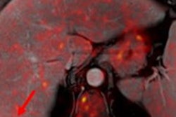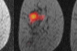
PET/MRI is better than MRI alone for the detection and staging of nodes and distant metastases in women with breast cancer, according to a study presented by German researchers at the recent RSNA 2019 meeting.
Researchers from the University of Düsseldorf in Germany found that the combination of PET with MRI was statistically superior to MRI alone for normal detection and staging, as well as in the detection of distant metastases (M staging).
"Both modalities have the diagnostic accuracy in detecting the M stage, but also bear the risk to overestimate the M stage," said study co-author and presenter Dr. Nils Bruckmann. "Therefore, PET/MRI should be considered as a useful alternative for the systematic staging of breast cancer patients at time of diagnosis."
Appropriate breast cancer treatment depends heavily on initial staging. Current guidelines recommend mammography, sonography, FDG-PET/CT, and breast MRI as effective methods to evaluate the local tumor extent, with the tumor, node, and metastases (TNM) system used to describe the spread of cancer.
"Consequently, FDG-PET/MRI could be an all-in-one solution involving dedicated breast MRI and PET," Bruckmann told RSNA attendees. "Therefore, the purpose of our study was to evaluate the diagnostic accuracy of whole-body MRI to whole-body FDG-PET/MRI for the initial TNM staging for newly diagnosed histologically proven breast cancer patients."
For this prospective, multicenter study, the researchers included 104 consecutive women (mean age, 53.4 ± 12.5 years) with histologically proven breast cancer. All subjects underwent whole-body FDG-PET/MRI in the supine position from the head to the midthigh using a 3-tesla MRI and PET system.
Two experienced radiologists separately analyzed the results on a per-patient and per-lesion basis and repeated the process four weeks later to avoid recognition bias. Their evaluations include the number of lesions, the lesion type, locations, sizes, and diagnostic confidence. Histopathology or follow-up exams were used as the reference standard, with that information available for all 104 patients.
A total of 298 lesions were included in the study. As for the nodal analysis, PET/MRI correctly detected nodes in more patients (83%) than MRI (74%), as well as more patients with positive nodes. PET/MRI also detected 87% of all nodes, compared with 75% for MRI alone. All of the aforementioned results were statistically significant in favor of PET/MRI.
| MRI vs. PET/MRI for staging breast cancer lesions | ||
| MRI alone | PET/MRI | |
| No. of patients | 74% | 83% |
| Nodal-positive patients | 62% | 75% |
| Detected lesions | 75% | 87% |
The researchers also noted distant metastases in seven patients (6%). PET/MRI identified all seven subjects (100%) and detected all of their metastases (100%). Unfortunately, both PET/MRI and MRI alone also had false-positive findings in four patients. Relative to the lesion-based analysis, PET/MRI detected all 31 metastatic lesions (100%). MRI detected only 18 (58%) of those metastases, resulting in a statistically significant difference in favor of PET/MRI.
"Our study demonstrates that PET/MRI has a superior diagnostic performance on a per-patient basis and on a lesion-based basis, compared with MRI alone in the TNM stage," Bruckmann said.


![Examples of ultrasound findings and techniques. (A) Images in a 39-year-old male patient with a mass in the left thigh. The mass is heterogeneous on the B-mode US image (compared with the patient in D) and showed increased microvascularity (superb microvascular imaging [SMI]) and shear-wave elastography (SWE) values. Undifferentiated pleomorphic sarcoma was diagnosed at biopsy (with pleomorphic rhabdomyosarcoma in surgical specimen). (B) Images in an 18-year-old male patient with a mass in the left leg. The mass is hypoechoic on the B-mode image, with no other findings suggestive of malignancy. The lesion is in contact with the cortex of the tibia, which is slightly irregular. CT revealed a doubtful anteromedial tibial erosion. The microvascular study demonstrated high vascularization, suggestive of malignancy. Periosteal Ewing sarcoma was diagnosed with both histologic and immunohistochemical confirmation. (C) Images in a 69-year-old female patient with a lump growing on the outside of the left leg. Multiple SWE examinations were performed (please note the high values obtained in the measurements, whereas the color map highlights the stiffness relative to adjacent tissues). SMI showed areas of increased vascularization to target for sampling. Undifferentiated spindle cell sarcoma was diagnosed at biopsy, with residual leiomyosarcoma in the surgical specimen after neoadjuvant therapy. (D) Images in a 56-year-old female patient with a mass in the right thigh. The mass is heterogeneous at both B-mode ultrasound (similar to patient A) and MRI (coronal T2-weighted spectral attenuated inversion recovery [SPAIR]; T1-weighted pre-contrast and postcontrast imaging), which even shows uptake after the administration of paramagnetic contrast material, which is traditionally suggestive of malignancy. Low values at SMI and elastography are suggestive of benignity. Spindle cell lipoma was diagnosed at biopsy, with atypical spindle cell lipomatous tumor in the surgical specimen.](https://img.auntminnieeurope.com/mindful/smg/workspaces/default/uploads/2026/08/images-radiol250278fig2.APCFLSvX6p.jpg?auto=format%2Ccompress&fit=crop&h=100&q=70&w=100)


![Examples of ultrasound findings and techniques. (A) Images in a 39-year-old male patient with a mass in the left thigh. The mass is heterogeneous on the B-mode US image (compared with the patient in D) and showed increased microvascularity (superb microvascular imaging [SMI]) and shear-wave elastography (SWE) values. Undifferentiated pleomorphic sarcoma was diagnosed at biopsy (with pleomorphic rhabdomyosarcoma in surgical specimen). (B) Images in an 18-year-old male patient with a mass in the left leg. The mass is hypoechoic on the B-mode image, with no other findings suggestive of malignancy. The lesion is in contact with the cortex of the tibia, which is slightly irregular. CT revealed a doubtful anteromedial tibial erosion. The microvascular study demonstrated high vascularization, suggestive of malignancy. Periosteal Ewing sarcoma was diagnosed with both histologic and immunohistochemical confirmation. (C) Images in a 69-year-old female patient with a lump growing on the outside of the left leg. Multiple SWE examinations were performed (please note the high values obtained in the measurements, whereas the color map highlights the stiffness relative to adjacent tissues). SMI showed areas of increased vascularization to target for sampling. Undifferentiated spindle cell sarcoma was diagnosed at biopsy, with residual leiomyosarcoma in the surgical specimen after neoadjuvant therapy. (D) Images in a 56-year-old female patient with a mass in the right thigh. The mass is heterogeneous at both B-mode ultrasound (similar to patient A) and MRI (coronal T2-weighted spectral attenuated inversion recovery [SPAIR]; T1-weighted pre-contrast and postcontrast imaging), which even shows uptake after the administration of paramagnetic contrast material, which is traditionally suggestive of malignancy. Low values at SMI and elastography are suggestive of benignity. Spindle cell lipoma was diagnosed at biopsy, with atypical spindle cell lipomatous tumor in the surgical specimen.](https://img.auntminnieeurope.com/mindful/smg/workspaces/default/uploads/2026/08/images-radiol250278fig2.APCFLSvX6p.jpg?auto=format%2Ccompress&fit=crop&h=112&q=70&w=112)