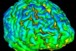
A Swedish-led group has developed a novel technique that uses florbetapir-PET scans for tracking beta-amyloid accumulation in adults to gauge the risk of progression to Alzheimer's disease. The investigators published their findings online on 17 July in JAMA Neurology.
By targeting certain brain regions and key biomarkers over time, the approach shows not only the degree of beta-amyloid involvement in the brain but also its growth and subsequent effect on cognitive decline, wrote the researchers led by Dr. Niklas Mattsson, PhD, in the Clinical Memory Research Unit at Lund University.
Beta-amyloid deposits have been linked to the onset of cognitive decline, mild cognitive impairment, and Alzheimer's disease, and they can affect different regions of the brain at various times -- making it possible to chart increases in accumulation over time in vivo. In fact, previous PET studies have shown that cerebrospinal fluid could be used as one biomarker to identify brain regions where beta-amyloid accumulation begins and areas that are affected later in the course of the disease.
"We used this method to construct a staging system, which we validated by examining the prevalence of unambiguously classified participants, differences in cognition by stage, biomarkers and imaging, regional gene expression, and longitudinal transition, because it is crucial to demonstrate that individuals are at risk for transitions from lower to higher stages," the authors explained.
The longitudinal multicenter study included 741 participants from the Alzheimer's Disease Neuroimaging Initiative (ADNI) who underwent at least two florbetapir-PET scans between June 2010 and July 2018. The study included a total of 2,072 PET scans in a population that included 304 subjects with no cognitive impairment, 384 with mild cognitive impairment, and 53 with Alzheimer's disease.
The researchers' methodology targeted cerebrospinal fluid and uptake of the PET radiopharmaceutical florbetapir (Amyvid, Eli Lilly) to identify early, intermediate, and late beta-amyloid accumulation in brain regions known to show that timing and location tendency. The beta-amyloid stages ranged from 0 to 3, correlating with the degree of beta-amyloid deposition. Florbetapir-PET scans were performed at baseline and again at two-, four-, and six-year follow-up.
As one might expect, most cognitively normal subjects -- 214 (70%), to be exact -- were in stage 0 at baseline. Interestingly, most participants with mild cognitive impairment were in either stage 0 (179, 47%) or stage 3 (135, 35%) of beta-amyloid deposition at baseline. Predictably, 42 Alzheimer's patients (79%) were characterized at stage 3.
Extrapolating those numbers to subsequent florbetapir-PET scans and beta-amyloid accumulation, the researchers figured that patients with stage 0 baseline results on PET scans had the lowest chance of advancing to a higher stage. Subjects with greater beta-amyloid accumulation and higher stage rating were at greater risk of their condition getting worse over time.
| Probability of advancing to higher stage rating based on beta-amyloid accumulation | ||
| At baseline | No. of patients | Probability |
| Stage 0 | 402 | 14.7% |
| Stage 1 | 21 | 71.4% |
| Stage 2 | 79 | 53.1% |
| Stage 3 | 227 | Not applicable |
As for this model's utility, Mattsson and colleagues suggest it might be "useful for early diagnosis, drug development, and to study disease mechanisms. Future studies can further investigate the regional vulnerability that underlies the stages."


![Examples of ultrasound findings and techniques. (A) Images in a 39-year-old male patient with a mass in the left thigh. The mass is heterogeneous on the B-mode US image (compared with the patient in D) and showed increased microvascularity (superb microvascular imaging [SMI]) and shear-wave elastography (SWE) values. Undifferentiated pleomorphic sarcoma was diagnosed at biopsy (with pleomorphic rhabdomyosarcoma in surgical specimen). (B) Images in an 18-year-old male patient with a mass in the left leg. The mass is hypoechoic on the B-mode image, with no other findings suggestive of malignancy. The lesion is in contact with the cortex of the tibia, which is slightly irregular. CT revealed a doubtful anteromedial tibial erosion. The microvascular study demonstrated high vascularization, suggestive of malignancy. Periosteal Ewing sarcoma was diagnosed with both histologic and immunohistochemical confirmation. (C) Images in a 69-year-old female patient with a lump growing on the outside of the left leg. Multiple SWE examinations were performed (please note the high values obtained in the measurements, whereas the color map highlights the stiffness relative to adjacent tissues). SMI showed areas of increased vascularization to target for sampling. Undifferentiated spindle cell sarcoma was diagnosed at biopsy, with residual leiomyosarcoma in the surgical specimen after neoadjuvant therapy. (D) Images in a 56-year-old female patient with a mass in the right thigh. The mass is heterogeneous at both B-mode ultrasound (similar to patient A) and MRI (coronal T2-weighted spectral attenuated inversion recovery [SPAIR]; T1-weighted pre-contrast and postcontrast imaging), which even shows uptake after the administration of paramagnetic contrast material, which is traditionally suggestive of malignancy. Low values at SMI and elastography are suggestive of benignity. Spindle cell lipoma was diagnosed at biopsy, with atypical spindle cell lipomatous tumor in the surgical specimen.](https://img.auntminnieeurope.com/mindful/smg/workspaces/default/uploads/2026/08/images-radiol250278fig2.APCFLSvX6p.jpg?auto=format%2Ccompress&fit=crop&h=100&q=70&w=100)


![Examples of ultrasound findings and techniques. (A) Images in a 39-year-old male patient with a mass in the left thigh. The mass is heterogeneous on the B-mode US image (compared with the patient in D) and showed increased microvascularity (superb microvascular imaging [SMI]) and shear-wave elastography (SWE) values. Undifferentiated pleomorphic sarcoma was diagnosed at biopsy (with pleomorphic rhabdomyosarcoma in surgical specimen). (B) Images in an 18-year-old male patient with a mass in the left leg. The mass is hypoechoic on the B-mode image, with no other findings suggestive of malignancy. The lesion is in contact with the cortex of the tibia, which is slightly irregular. CT revealed a doubtful anteromedial tibial erosion. The microvascular study demonstrated high vascularization, suggestive of malignancy. Periosteal Ewing sarcoma was diagnosed with both histologic and immunohistochemical confirmation. (C) Images in a 69-year-old female patient with a lump growing on the outside of the left leg. Multiple SWE examinations were performed (please note the high values obtained in the measurements, whereas the color map highlights the stiffness relative to adjacent tissues). SMI showed areas of increased vascularization to target for sampling. Undifferentiated spindle cell sarcoma was diagnosed at biopsy, with residual leiomyosarcoma in the surgical specimen after neoadjuvant therapy. (D) Images in a 56-year-old female patient with a mass in the right thigh. The mass is heterogeneous at both B-mode ultrasound (similar to patient A) and MRI (coronal T2-weighted spectral attenuated inversion recovery [SPAIR]; T1-weighted pre-contrast and postcontrast imaging), which even shows uptake after the administration of paramagnetic contrast material, which is traditionally suggestive of malignancy. Low values at SMI and elastography are suggestive of benignity. Spindle cell lipoma was diagnosed at biopsy, with atypical spindle cell lipomatous tumor in the surgical specimen.](https://img.auntminnieeurope.com/mindful/smg/workspaces/default/uploads/2026/08/images-radiol250278fig2.APCFLSvX6p.jpg?auto=format%2Ccompress&fit=crop&h=112&q=70&w=112)