Korean researchers have found that contrast-enhanced digital mammography (CEDM) works just as well as contrast-enhanced breast MRI (CE-MRI) for measuring breast tumors before surgery, according to a study published on 24 April in the British Journal of Radiology.
Preoperative breast imaging to measure tumor size helps clinicians determine the extent of disease and is a key component of surgical planning, wrote a group led by Dr. Inyoung Youn, PhD, of Sungkyunkwan University in Seoul. MRI is the most widely used imaging modality for this application and is considered to be the most accurate.
But having another option would give surgeons a wider range of tools and address some of breast MRI's limitations.
"Breast MRI is an important imaging modality for breast cancer patients; however, the high cost of the procedure, claustrophobia, longer study time, and lesser accessibility ... compared with mammography are a few of its limitations," the team wrote.
Previous clinical research has compared the diagnostic performance of CEDM and CE-MRI and found that the two modalities are comparable in their accuracy; however, these studies haven't compared their performance in measuring tumor size before surgery. So Youn and colleagues compared the accuracy of CEDM and MRI, including maximum intensity projection (MIP) images, for measuring histologically confirmed breast tumors.
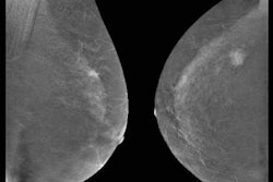
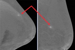
The study included 52 women with 54 cancers who had undergone contrast-enhanced MRI and contrast-enhanced digital mammography within a week before surgery for breast cancer between November 2016 and March 2017. The researchers assessed the two modalities' performance by comparing the maximum diameter of the main lesion on mediolateral oblique (MLO) and craniocaudal (CC) digital mammography and CEDM with sagittal and axial MIP images and dynamic CE-MRI before surgery. No patients experienced side effects from contrast media.
Of the 54 cancers, 41 were treated with breast-conserving surgery and 13 were treated with mastectomy. Mean lesion size was 15.5 mm. All parameters of CEDM and CE-MRI showed good agreement (kappa > 0.75), the group found.
| CEDM vs. CE-MRI for measuring mean breast tumor size | |||
| Mammography | CEDM | CE-MRI | |
| MLO | 21.1 mm | 20.3 mm | -- |
| CC | 21.2 mm | 20 mm | -- |
| Sagittal MIP | -- | -- | 19.8 mm |
| Axial MIP | -- | -- | 17.7 mm |
The researchers also discovered that both modalities visualized the size of the tumor more accurately in the axial plane than the sagittal, and that the CC view on CEDM was comparable or superior to that of CE-MRI.
Because CEDM and CE-MRI perform comparably when it comes to measuring breast lesions, clinicians have more options, according to the group.
"CEDM can be used as an alternative modality to CE-MRI in tumor size measurement, and the axial plane of the CEDM and CE-MRI would be the first choice for image review and surgical planning," the team concluded.


![Examples of ultrasound findings and techniques. (A) Images in a 39-year-old male patient with a mass in the left thigh. The mass is heterogeneous on the B-mode US image (compared with the patient in D) and showed increased microvascularity (superb microvascular imaging [SMI]) and shear-wave elastography (SWE) values. Undifferentiated pleomorphic sarcoma was diagnosed at biopsy (with pleomorphic rhabdomyosarcoma in surgical specimen). (B) Images in an 18-year-old male patient with a mass in the left leg. The mass is hypoechoic on the B-mode image, with no other findings suggestive of malignancy. The lesion is in contact with the cortex of the tibia, which is slightly irregular. CT revealed a doubtful anteromedial tibial erosion. The microvascular study demonstrated high vascularization, suggestive of malignancy. Periosteal Ewing sarcoma was diagnosed with both histologic and immunohistochemical confirmation. (C) Images in a 69-year-old female patient with a lump growing on the outside of the left leg. Multiple SWE examinations were performed (please note the high values obtained in the measurements, whereas the color map highlights the stiffness relative to adjacent tissues). SMI showed areas of increased vascularization to target for sampling. Undifferentiated spindle cell sarcoma was diagnosed at biopsy, with residual leiomyosarcoma in the surgical specimen after neoadjuvant therapy. (D) Images in a 56-year-old female patient with a mass in the right thigh. The mass is heterogeneous at both B-mode ultrasound (similar to patient A) and MRI (coronal T2-weighted spectral attenuated inversion recovery [SPAIR]; T1-weighted pre-contrast and postcontrast imaging), which even shows uptake after the administration of paramagnetic contrast material, which is traditionally suggestive of malignancy. Low values at SMI and elastography are suggestive of benignity. Spindle cell lipoma was diagnosed at biopsy, with atypical spindle cell lipomatous tumor in the surgical specimen.](https://img.auntminnieeurope.com/mindful/smg/workspaces/default/uploads/2026/08/images-radiol250278fig2.APCFLSvX6p.jpg?auto=format%2Ccompress&fit=crop&h=100&q=70&w=100)


![Examples of ultrasound findings and techniques. (A) Images in a 39-year-old male patient with a mass in the left thigh. The mass is heterogeneous on the B-mode US image (compared with the patient in D) and showed increased microvascularity (superb microvascular imaging [SMI]) and shear-wave elastography (SWE) values. Undifferentiated pleomorphic sarcoma was diagnosed at biopsy (with pleomorphic rhabdomyosarcoma in surgical specimen). (B) Images in an 18-year-old male patient with a mass in the left leg. The mass is hypoechoic on the B-mode image, with no other findings suggestive of malignancy. The lesion is in contact with the cortex of the tibia, which is slightly irregular. CT revealed a doubtful anteromedial tibial erosion. The microvascular study demonstrated high vascularization, suggestive of malignancy. Periosteal Ewing sarcoma was diagnosed with both histologic and immunohistochemical confirmation. (C) Images in a 69-year-old female patient with a lump growing on the outside of the left leg. Multiple SWE examinations were performed (please note the high values obtained in the measurements, whereas the color map highlights the stiffness relative to adjacent tissues). SMI showed areas of increased vascularization to target for sampling. Undifferentiated spindle cell sarcoma was diagnosed at biopsy, with residual leiomyosarcoma in the surgical specimen after neoadjuvant therapy. (D) Images in a 56-year-old female patient with a mass in the right thigh. The mass is heterogeneous at both B-mode ultrasound (similar to patient A) and MRI (coronal T2-weighted spectral attenuated inversion recovery [SPAIR]; T1-weighted pre-contrast and postcontrast imaging), which even shows uptake after the administration of paramagnetic contrast material, which is traditionally suggestive of malignancy. Low values at SMI and elastography are suggestive of benignity. Spindle cell lipoma was diagnosed at biopsy, with atypical spindle cell lipomatous tumor in the surgical specimen.](https://img.auntminnieeurope.com/mindful/smg/workspaces/default/uploads/2026/08/images-radiol250278fig2.APCFLSvX6p.jpg?auto=format%2Ccompress&fit=crop&h=112&q=70&w=112)