Researchers from Spain have used a 3D-printed model of the thoracic wall to plan and guide tumor resection for a 64-year-old man -- allowing them to complete the surgery two hours faster than the average operating time.
The group from Biodonostia Health Research Institute in Spain created the 3D-printed model to help resolve the case of a tumor that was lodged in the thoracic wall. To remove the tumor, a team of surgeons faced the challenge of removing more than one of the patient's ribs without damaging the lungs, according to a report from Stratasys.
Due to the complexity of the procedure, the researchers decided to create an individually tailored 3D-printed model of the patient's thoracic wall and tumor for the surgeons. They did so by converting the patient's CT scans into 3D virtual models and then sending them to a 3D printer (Fortus 450mc, Stratasys) that produced the model in less than 24 hours.
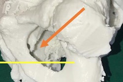
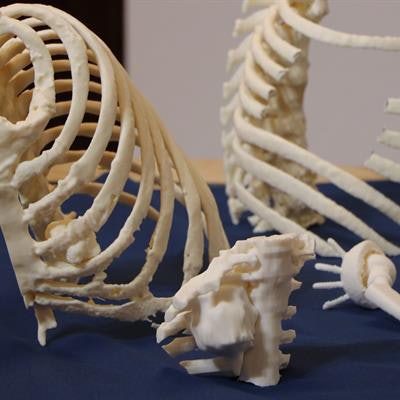
 3D-printed thoracic wall and tumors used as a surgical guide. Image courtesy of Stratasys.
Examining the model during presurgical planning allowed the surgical team to determine the type of screws and properly shape the titanium plates that the patient would require even before commencing surgery. This resulted in reducing operating time by roughly two hours, compared with the hospital average.
In addition, the group used the 3D-printed model to explain to the patient the nature of his condition and the intended course of treatment. The surgical consult ended up being more efficient than usual, according to Dr. Jon Zabaleta, one of the thoracic surgeons for the case.
"The use of the 3D-printed model was so essential to this case, and we are working to apply this to many other surgical disciplines across the hospital, from pancreatic tumors to airway stenosis, and these 3D-printed models are already being used to help train our future surgeons," he noted in the Stratasys statement.