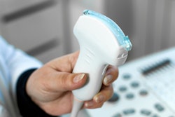
2D shear-wave elastography (SWE) is an effective tool for ruling out liver fibrosis or cirrhosis in people with chronic liver disease, German researchers wrote in a study published online on 11 December in the Journal of Ultrasound in Medicine.
The findings suggest a less-invasive alternative for these patients, wrote the team led by Dr. Golo Petzold from University Medical Center Göttingen.
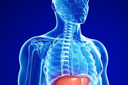
"In the treatment and monitoring of patients with chronic liver disease, the diagnosis of liver fibrosis or cirrhosis is of great importance," the researchers wrote. "The detection of liver cirrhosis can be challenging at times; early forms of cirrhosis and advanced fibrosis are especially hard to distinguish by imaging studies. The reference standard for the diagnosis of liver fibrosis (staging) and cirrhosis is liver biopsy. This procedure is invasive, is sometimes painful, and has rare but potentially serious complications."
An increasing number of ultrasound device manufacturers are incorporating shear-wave elastography into their scanners, according to Petzold and colleagues. Their study had three goals:
- Use 2D SWE to determine normal liver stiffness values for healthy subjects and compare them with values in patients with liver disease.
- Assess the influence of age, sex, body mass index, and steatosis on liver stiffness measurement.
- Explore whether it's possible to rule out disease based on cutoff values for histologically confirmed cirrhosis.
The study included 167 subjects enrolled between July 2016 and February 2018. Within this group, 56 had a healthy liver but nonhepatic disease (requiring baseline abdominal ultrasound), 45 had liver cirrhosis, and 66 were healthy volunteers. All participants underwent a baseline 2D SWE ultrasound exam to establish a liver stiffness measurement.
Liver stiffness in the patients with healthy livers but nonhepatic disease did not differ significantly from liver stiffness in the healthy volunteers, the researchers found. Male sex was associated with significantly higher liver stiffness in healthy volunteers; however, age, body mass index, mild steatosis, and nonhepatic morbidities had no significant impact on liver stiffness. Liver stiffness was significantly higher in patients with cirrhosis.
| Liver stiffness values on SWE by patient group | |||
| Healthy participants | Healthy liver patients with nonhepatic disease | Patients with cirrhosis | |
| Liver stiffness (kPa) | 5.19 | 4.93 | 13.29 |
| Minimum liver stiffness (kPa) | 3.62 | 3.49 | 7.38 |
| Maximum liver stiffness (kPa) | 8.33 | 6.66 | 19.96 |
2D SWE is a relatively new ultrasound technique, but it appears to be effective for evaluating liver stiffness and, therefore, ruling out cirrhosis, Petzold and colleagues wrote.
"The liver stiffness measurement in patients with histologically confirmed liver cirrhosis was significantly higher than that in the healthy-liver cohort," they concluded. "Importantly, the reference ranges for the cirrhosis and healthy-liver cohorts did not overlap. ... Therefore, on the basis of our data, we can rule out cirrhosis in patients with normal liver stiffness values, but no cutoff values for cirrhosis can be derived at present."