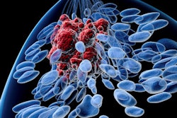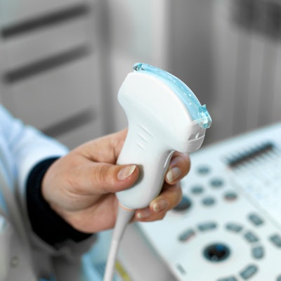
A 3D-printed device with built-in pressure sensors that fits around an ultrasound transducer like a shell could produce more accurate shear-wave elastography (SWE) measurements for breast imaging and other use cases. Researchers describe the prototype attachment in a 15 June article in Ultrasound in Medicine & Biology.
Applying too much pressure during a handheld ultrasound scan can affect the accuracy of tissue elasticity measurements. The prototype device helps to overcome this limitation by using a series of interlocking plastic shells that measure pressure changes in real-time. With further refinements, it could one day help radiologists apply consistent, minimal pressure.
"Shear-wave elastography may produce misleadingly high values if too much pressure is applied during the imaging process," wrote the authors, led by Katrin Skerl, PhD, a professor of medical and mechanical engineering at Furtwangen University in Germany. "However, in clinical routine, there is presently no way to monitor the pressure applied to an ultrasonic imaging transducer and allows observation of the applied pressure in real time."
For the device, Skerl and colleagues used cheap, 3D-printed plastic parts to create layers that surround an ultrasound SWE transducer. The device contains two plastic shells: an inner layer with pressure sensors and an outer layer with independent, sliding movement.
The inner layer attaches directly to the transducer, and springs push the outer layer upward onto the inner layers' sensors. As the outer layer slides during a scan, the inner sensors calculate the associated reduction in pressure to provide real-time feedback.
The researchers validated the prototype with the help of two radiologists and a radiographer experienced in breast sonography and elastography. The breast imagers used the device to scan chicken breast, porcine belly, beef, and bovine udder samples resting on top of a calibrated scale.
Each imager began by applying the minimal amount of pressure possible, then increased pressure until the image quality was insufficient, the sample was damaged, or SWE measurements reached 10 Newtons (N) or 20 kilopascals (kPa), an upper threshold set by an ultrasound breast imaging radiologist with more than 20 years of clinical experience. During the experiment, the mean elasticity measurement increased with pressure, validating the probe's ability to provide real-time feedback.
The prototype is an exciting first step toward creating a device to monitor real-time pressure during handheld ultrasound scans, and it was of the first prototypes designed to work for breast imaging. But to be clinically relevant, newer models will need to be made from higher quality material, account for pressure from the weight of the probe and the cable, and be easier to add and remove, the authors noted.
The benefits of such a device could be game-changing for sonographers performing shear-wave elastography, according to the authors. Not only could it reduce pressure bias in elasticity measurements and give radiologists real-time feedback but it could also pave the way for creating protocols with pressure guidelines for optimal performance, or even protocols with ideal pressure ranges for different tumor types.
"The measurement device introduced here is the first step toward introducing a method for examining the pressure applied during clinical examinations," the authors concluded. "The results from a preliminary ex vivo study showing approximate linear increase in elasticity are promising. However, the device design should be improved to enhance its clinical applicability."


![Examples of ultrasound findings and techniques. (A) Images in a 39-year-old male patient with a mass in the left thigh. The mass is heterogeneous on the B-mode US image (compared with the patient in D) and showed increased microvascularity (superb microvascular imaging [SMI]) and shear-wave elastography (SWE) values. Undifferentiated pleomorphic sarcoma was diagnosed at biopsy (with pleomorphic rhabdomyosarcoma in surgical specimen). (B) Images in an 18-year-old male patient with a mass in the left leg. The mass is hypoechoic on the B-mode image, with no other findings suggestive of malignancy. The lesion is in contact with the cortex of the tibia, which is slightly irregular. CT revealed a doubtful anteromedial tibial erosion. The microvascular study demonstrated high vascularization, suggestive of malignancy. Periosteal Ewing sarcoma was diagnosed with both histologic and immunohistochemical confirmation. (C) Images in a 69-year-old female patient with a lump growing on the outside of the left leg. Multiple SWE examinations were performed (please note the high values obtained in the measurements, whereas the color map highlights the stiffness relative to adjacent tissues). SMI showed areas of increased vascularization to target for sampling. Undifferentiated spindle cell sarcoma was diagnosed at biopsy, with residual leiomyosarcoma in the surgical specimen after neoadjuvant therapy. (D) Images in a 56-year-old female patient with a mass in the right thigh. The mass is heterogeneous at both B-mode ultrasound (similar to patient A) and MRI (coronal T2-weighted spectral attenuated inversion recovery [SPAIR]; T1-weighted pre-contrast and postcontrast imaging), which even shows uptake after the administration of paramagnetic contrast material, which is traditionally suggestive of malignancy. Low values at SMI and elastography are suggestive of benignity. Spindle cell lipoma was diagnosed at biopsy, with atypical spindle cell lipomatous tumor in the surgical specimen.](https://img.auntminnieeurope.com/mindful/smg/workspaces/default/uploads/2026/08/images-radiol250278fig2.APCFLSvX6p.jpg?auto=format%2Ccompress&fit=crop&h=100&q=70&w=100)


![Examples of ultrasound findings and techniques. (A) Images in a 39-year-old male patient with a mass in the left thigh. The mass is heterogeneous on the B-mode US image (compared with the patient in D) and showed increased microvascularity (superb microvascular imaging [SMI]) and shear-wave elastography (SWE) values. Undifferentiated pleomorphic sarcoma was diagnosed at biopsy (with pleomorphic rhabdomyosarcoma in surgical specimen). (B) Images in an 18-year-old male patient with a mass in the left leg. The mass is hypoechoic on the B-mode image, with no other findings suggestive of malignancy. The lesion is in contact with the cortex of the tibia, which is slightly irregular. CT revealed a doubtful anteromedial tibial erosion. The microvascular study demonstrated high vascularization, suggestive of malignancy. Periosteal Ewing sarcoma was diagnosed with both histologic and immunohistochemical confirmation. (C) Images in a 69-year-old female patient with a lump growing on the outside of the left leg. Multiple SWE examinations were performed (please note the high values obtained in the measurements, whereas the color map highlights the stiffness relative to adjacent tissues). SMI showed areas of increased vascularization to target for sampling. Undifferentiated spindle cell sarcoma was diagnosed at biopsy, with residual leiomyosarcoma in the surgical specimen after neoadjuvant therapy. (D) Images in a 56-year-old female patient with a mass in the right thigh. The mass is heterogeneous at both B-mode ultrasound (similar to patient A) and MRI (coronal T2-weighted spectral attenuated inversion recovery [SPAIR]; T1-weighted pre-contrast and postcontrast imaging), which even shows uptake after the administration of paramagnetic contrast material, which is traditionally suggestive of malignancy. Low values at SMI and elastography are suggestive of benignity. Spindle cell lipoma was diagnosed at biopsy, with atypical spindle cell lipomatous tumor in the surgical specimen.](https://img.auntminnieeurope.com/mindful/smg/workspaces/default/uploads/2026/08/images-radiol250278fig2.APCFLSvX6p.jpg?auto=format%2Ccompress&fit=crop&h=112&q=70&w=112)