Dear AuntMinnieEurope Member,
Brexit looks certain to have major implications for the practice of radiology and nuclear medicine in the U.K., and it's likely to have at least some effect on the rest of Europe too. The main difficulty, though, is to work out exactly what impact it will have.
More than 18 months have passed since market expert Stephen Holloway last wrote for us about this subject. In a new column, he summarizes the current situation, describes what's changed since early 2017, and speculates a little about the future. Visit the Molecular Imaging Community, or click here.
A large German-led study that looked at radiation dose administered to patients undergoing coronary CT angiography at 61 hospitals in 32 different countries during one month in 2017 has found the dose fell sharply compared with a similar survey 10 years earlier. This outcome is much better than the researchers expected. For the full details, go to the CT Community, or click here.
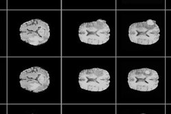
Artificial intelligence (AI) was a hot topic at last week's International Conference on Medical Image Computing and Computer Assisted Intervention in Granada, Spain. At the meeting, researchers explained how they've trained a type of AI algorithm to generate synthetic abnormal brain MR images. Find out more here.
Meanwhile, it's been a hectic few days in the world of breast imaging. The National Health Service Scotland is contacting nearly 1,800 women following revelations they did not receive invitations to their final round of breast screening. Head to the Women's Imaging Community, or click here.
Also, two vociferous opponents of breast screening, Dr. Peter Gøtzsche and Dr. H. Gilbert Welch, have lost positions in the last week. In Gøtzsche's case, he was removed from the board of the Cochrane group in a controversial action taken at its annual meeting in Edinburgh, Scotland. The change threatens to create a split within Cochrane as proponents of evidence-based medicine take sides. Get the story here.
Finally, a Scandinavian team is reviving the controversial claim that the recent drop in breast cancer mortality is not due to mammography screening but rather the result of improved treatment. The authors say it's time to consider alternatives to mammography screening, but is their recommendation of breast palpation by physicians a realistic option? Click here to learn more.


![Examples of ultrasound findings and techniques. (A) Images in a 39-year-old male patient with a mass in the left thigh. The mass is heterogeneous on the B-mode US image (compared with the patient in D) and showed increased microvascularity (superb microvascular imaging [SMI]) and shear-wave elastography (SWE) values. Undifferentiated pleomorphic sarcoma was diagnosed at biopsy (with pleomorphic rhabdomyosarcoma in surgical specimen). (B) Images in an 18-year-old male patient with a mass in the left leg. The mass is hypoechoic on the B-mode image, with no other findings suggestive of malignancy. The lesion is in contact with the cortex of the tibia, which is slightly irregular. CT revealed a doubtful anteromedial tibial erosion. The microvascular study demonstrated high vascularization, suggestive of malignancy. Periosteal Ewing sarcoma was diagnosed with both histologic and immunohistochemical confirmation. (C) Images in a 69-year-old female patient with a lump growing on the outside of the left leg. Multiple SWE examinations were performed (please note the high values obtained in the measurements, whereas the color map highlights the stiffness relative to adjacent tissues). SMI showed areas of increased vascularization to target for sampling. Undifferentiated spindle cell sarcoma was diagnosed at biopsy, with residual leiomyosarcoma in the surgical specimen after neoadjuvant therapy. (D) Images in a 56-year-old female patient with a mass in the right thigh. The mass is heterogeneous at both B-mode ultrasound (similar to patient A) and MRI (coronal T2-weighted spectral attenuated inversion recovery [SPAIR]; T1-weighted pre-contrast and postcontrast imaging), which even shows uptake after the administration of paramagnetic contrast material, which is traditionally suggestive of malignancy. Low values at SMI and elastography are suggestive of benignity. Spindle cell lipoma was diagnosed at biopsy, with atypical spindle cell lipomatous tumor in the surgical specimen.](https://img.auntminnieeurope.com/mindful/smg/workspaces/default/uploads/2026/08/images-radiol250278fig2.APCFLSvX6p.jpg?auto=format%2Ccompress&fit=crop&h=100&q=70&w=100)


![Examples of ultrasound findings and techniques. (A) Images in a 39-year-old male patient with a mass in the left thigh. The mass is heterogeneous on the B-mode US image (compared with the patient in D) and showed increased microvascularity (superb microvascular imaging [SMI]) and shear-wave elastography (SWE) values. Undifferentiated pleomorphic sarcoma was diagnosed at biopsy (with pleomorphic rhabdomyosarcoma in surgical specimen). (B) Images in an 18-year-old male patient with a mass in the left leg. The mass is hypoechoic on the B-mode image, with no other findings suggestive of malignancy. The lesion is in contact with the cortex of the tibia, which is slightly irregular. CT revealed a doubtful anteromedial tibial erosion. The microvascular study demonstrated high vascularization, suggestive of malignancy. Periosteal Ewing sarcoma was diagnosed with both histologic and immunohistochemical confirmation. (C) Images in a 69-year-old female patient with a lump growing on the outside of the left leg. Multiple SWE examinations were performed (please note the high values obtained in the measurements, whereas the color map highlights the stiffness relative to adjacent tissues). SMI showed areas of increased vascularization to target for sampling. Undifferentiated spindle cell sarcoma was diagnosed at biopsy, with residual leiomyosarcoma in the surgical specimen after neoadjuvant therapy. (D) Images in a 56-year-old female patient with a mass in the right thigh. The mass is heterogeneous at both B-mode ultrasound (similar to patient A) and MRI (coronal T2-weighted spectral attenuated inversion recovery [SPAIR]; T1-weighted pre-contrast and postcontrast imaging), which even shows uptake after the administration of paramagnetic contrast material, which is traditionally suggestive of malignancy. Low values at SMI and elastography are suggestive of benignity. Spindle cell lipoma was diagnosed at biopsy, with atypical spindle cell lipomatous tumor in the surgical specimen.](https://img.auntminnieeurope.com/mindful/smg/workspaces/default/uploads/2026/08/images-radiol250278fig2.APCFLSvX6p.jpg?auto=format%2Ccompress&fit=crop&h=112&q=70&w=112)