Dear Advanced Visualization Insider,
Last week's Computer Assisted Radiology and Surgery (CARS) 2018 international congress in Berlin featured a wide range of advancements in radiological imaging, including the continued integration of augmented reality into healthcare.
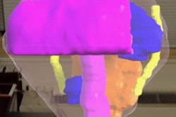
In one presentation at the meeting, a group of researchers from the New York University School of Medicine detailed their experience using an augmented reality device to visualize holographic models during presurgical planning for prostate cancer removal. Discover how well it fared compared with conventional MRI by clicking here.
Also at CARS 2018, researchers from the Netherlands demonstrated how their new technique for quantifying wrist motion using 4D CT could aid clinicians in the evaluation of bone displacement and fracture healing.
Clinicians have been referring to 3D models to examine anatomy and pathology increasingly more often in recent years. A prime example was the use of volume-rendered CT scans to reveal a selection of striking injuries at the running of the bulls at the Fiesta de San Fermín in Pamplona, Spain, last year.
Taking 3D imaging one step further, a team of investigators from the Fraunhofer Institute for Medical Image Computing (MEVIS) in Bremen, Germany, have developed artificial intelligence algorithms capable of creating 3D models based on MRI or CT scans of the liver. A separate team designed a joint space mapping technique that uses algorithms to determine the space between joint bones on 3D CT scans.
Applying 3D imaging to brain visualization, researchers from Belgium combined biophysics and functional MRI to construct personalized virtual brain models. Learn what these models uncovered about brain function in patients with brain tumors by clicking here.
In other news, Ulrik Kristensen, a senior market analyst at Signify Research, explored the hype surrounding cloud technology and its potential for advanced visualization. Find out why it's taking so long for the technology to materialize by clicking here.
Finally, the ongoing application of 3D printing in medicine has made considerable headway in improving patient education for those with brain tumors, kidney transplantation, and CT radiation dose management.
Check out more related stories and news in the Advanced Visualization Community at AuntMinnieEurope.com.


![Examples of ultrasound findings and techniques. (A) Images in a 39-year-old male patient with a mass in the left thigh. The mass is heterogeneous on the B-mode US image (compared with the patient in D) and showed increased microvascularity (superb microvascular imaging [SMI]) and shear-wave elastography (SWE) values. Undifferentiated pleomorphic sarcoma was diagnosed at biopsy (with pleomorphic rhabdomyosarcoma in surgical specimen). (B) Images in an 18-year-old male patient with a mass in the left leg. The mass is hypoechoic on the B-mode image, with no other findings suggestive of malignancy. The lesion is in contact with the cortex of the tibia, which is slightly irregular. CT revealed a doubtful anteromedial tibial erosion. The microvascular study demonstrated high vascularization, suggestive of malignancy. Periosteal Ewing sarcoma was diagnosed with both histologic and immunohistochemical confirmation. (C) Images in a 69-year-old female patient with a lump growing on the outside of the left leg. Multiple SWE examinations were performed (please note the high values obtained in the measurements, whereas the color map highlights the stiffness relative to adjacent tissues). SMI showed areas of increased vascularization to target for sampling. Undifferentiated spindle cell sarcoma was diagnosed at biopsy, with residual leiomyosarcoma in the surgical specimen after neoadjuvant therapy. (D) Images in a 56-year-old female patient with a mass in the right thigh. The mass is heterogeneous at both B-mode ultrasound (similar to patient A) and MRI (coronal T2-weighted spectral attenuated inversion recovery [SPAIR]; T1-weighted pre-contrast and postcontrast imaging), which even shows uptake after the administration of paramagnetic contrast material, which is traditionally suggestive of malignancy. Low values at SMI and elastography are suggestive of benignity. Spindle cell lipoma was diagnosed at biopsy, with atypical spindle cell lipomatous tumor in the surgical specimen.](https://img.auntminnieeurope.com/mindful/smg/workspaces/default/uploads/2026/08/images-radiol250278fig2.APCFLSvX6p.jpg?auto=format%2Ccompress&fit=crop&h=100&q=70&w=100)


![Examples of ultrasound findings and techniques. (A) Images in a 39-year-old male patient with a mass in the left thigh. The mass is heterogeneous on the B-mode US image (compared with the patient in D) and showed increased microvascularity (superb microvascular imaging [SMI]) and shear-wave elastography (SWE) values. Undifferentiated pleomorphic sarcoma was diagnosed at biopsy (with pleomorphic rhabdomyosarcoma in surgical specimen). (B) Images in an 18-year-old male patient with a mass in the left leg. The mass is hypoechoic on the B-mode image, with no other findings suggestive of malignancy. The lesion is in contact with the cortex of the tibia, which is slightly irregular. CT revealed a doubtful anteromedial tibial erosion. The microvascular study demonstrated high vascularization, suggestive of malignancy. Periosteal Ewing sarcoma was diagnosed with both histologic and immunohistochemical confirmation. (C) Images in a 69-year-old female patient with a lump growing on the outside of the left leg. Multiple SWE examinations were performed (please note the high values obtained in the measurements, whereas the color map highlights the stiffness relative to adjacent tissues). SMI showed areas of increased vascularization to target for sampling. Undifferentiated spindle cell sarcoma was diagnosed at biopsy, with residual leiomyosarcoma in the surgical specimen after neoadjuvant therapy. (D) Images in a 56-year-old female patient with a mass in the right thigh. The mass is heterogeneous at both B-mode ultrasound (similar to patient A) and MRI (coronal T2-weighted spectral attenuated inversion recovery [SPAIR]; T1-weighted pre-contrast and postcontrast imaging), which even shows uptake after the administration of paramagnetic contrast material, which is traditionally suggestive of malignancy. Low values at SMI and elastography are suggestive of benignity. Spindle cell lipoma was diagnosed at biopsy, with atypical spindle cell lipomatous tumor in the surgical specimen.](https://img.auntminnieeurope.com/mindful/smg/workspaces/default/uploads/2026/08/images-radiol250278fig2.APCFLSvX6p.jpg?auto=format%2Ccompress&fit=crop&h=112&q=70&w=112)