Researchers from Belgium are questioning whether organ-based tube-current modulation in CT chest studies of women really reduces radiation dose. They found that while radiation dose to the breast fell, it increased in other areas, according to a study published on March 27 in Radiology.
In the study based on computer simulations, the group from Ghent University created customized 3D voxel models of the chest, abdomen, and pelvis from the scans of patients who underwent a conventional CT protocol as well as an organ-based tube-current modulation protocol. Though the organ-based tube-current modulation software reduced radiation dose to the breast, it ended up increasing overall radiation dose to the body -- especially in the lungs, liver, and kidneys -- along with the cancer mortality rate, based on statistical models.
"The potential benefit of organ-based tube-current modulation to the female breast in chest CT is overestimated because the reduced tube current zone is too limited," first author Caro Franck and colleagues wrote. "[The] posterior organs will absorb up to 26% more radiation, resulting in no overall benefit for radiation-induced cancer risks."
Organ-based dose reduction
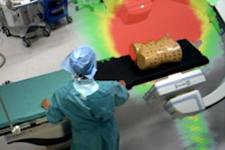
Due to the higher sensitivity of the breasts to radiation exposure, clinicians have been attempting to minimize radiation dose to the area on imaging exams using techniques such as organ-based tube-current modulation, according to the authors. This technique can reduce exposure to a designated 80° range on the anterior surface of the patient but at the cost of a compensatory increase in exposure to other areas (Radiology, March 27, 2018).
Prior research using organ-based tube-current modulations on CT phantoms has reported dose reductions to the breast ranging from 16% to 50%, they noted. However, these studies did not take into account the realistic clinical positioning of the breasts, which, in most cases, would have extended slightly beyond the indicated dose-reduction region.
Hoping to provide a more precise and comprehensive evaluation of organ-based tube-current modulation, the researchers evaluated the standard chest, abdominal, and pelvic CT scans of 50 female patients as well 17 other scans of these same patients in which organ-based tube-current modulation was used. Clinicians acquired the modified scans using an algorithm known as X-Care that is part of CT scanner tube-current modulation software (Care Dose4D, Siemens Healthineers). The algorithm allows users to set the same exposure parameters for scanning as conventional CT while also enabling breast-based tube-current modulation.
Next, the researchers created patient-specific 3D voxel models based on both the standard CT scans and tube-current modulated CT scans of each patient. By inputting these models into dose calculation software (ImpactMC 1.6, CT Imaging), they were able to perform three sets of Monte Carlo simulations to estimate radiation dose distributions for specific organs.
The simulations utilized the standard CT protocol models, the organ-based tube-current modulation models, or virtual models that modified the standard CT model by factoring in angular (x-y axis) modulation. The virtual CT models allowed the investigators to determine dose reductions associated with tube-current modulation theoretically, although these measurements ended up overestimating actual clinical dose reductions.
Increased cancer mortality rate
Comparing the results of the simulations, the group discovered that applying the organ-based tube-current modulation technique reduced radiation dose to the breast by 9% and the thyroid by 18%. However, there was an increase in average radiation dose to the lungs by 17%, the liver by 11%, and the kidneys by 26%.
| Chest CT radiation dose with and without organ-based tube-current modulation | |||
| Organ | Dose with standard CT (mGy) | Dose with organ-based tube-current modulation (mGy) | p-value |
| Thyroid | 21 | 17 | 0.04 |
| Breast | 17 | 16 | 0.003 |
| Lung | 15 | 17 | 0.005 |
| Liver | 15 | 16 | 0.01 |
| Kidney | 13 | 16 | < 0.001 |
Despite lowering radiation dose in the thyroid and breasts, using organ-based tube-current modulation led to a 15% increase in overall CT dose index volume (CTDIvol) values.
"Dose reductions to superficial and anteriorly located organs are still observed, but to a lesser extent [than the virtual model]," the researchers wrote. "Doses to other relevant organs in the field-of-view were higher with use of X-Care."
In terms of cancer risk, the overall increase in patient radiation exposure from organ-based tube-current modulation resulted in no significant change in lifetime attributable risk (LAR) for cancer incidence compared with conventional CT (p = 0.06), but it did lead to a statistically significant increase in LAR of cancer mortality (p = 0.006).
Subsequent image quality analysis of the CT scans revealed that the organ-based tube-current modulation protocol improved image resolution by an average of 7% in terms of noise parameters (p < 0.002).
This higher image quality suggests that there is potential to lower radiation exposure from the organ-based tube-current modulation protocol for future studies while still matching the image quality of standard CT scans, they noted. The extent to which this altered protocol affects radiation exposure and cancer mortality may shed more light on the utility of organ-based tube-current modulation.