The majority of CT exams ordered for patients with head trauma in the emergency room of an Italian hospital did not meet the appropriate referral criteria, according to research to be presented at the upcoming 2018 American Roentgen Ray Society (ARRS) meeting in Washington, DC.
The study, to be presented by Dr. Michaela Cellina from Fatebenefratelli-Sacco Hospital in Milan, reviewed the head CT scans of emergency room patients who presented with a minor head injury between January and June 2016 at the hospital. Among the 492 cases, 260 (52.8%) did not meet the National Institute for Health and Care Excellence (NICE) criteria for ordering a CT exam, and 376 (76.4%) did not meet the Canadian CT Head Rule (CCHR) criteria.
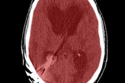
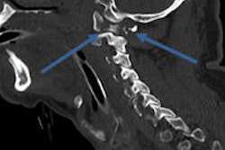
They found no statistically significant association between either physician specialty or experience and unwarranted referrals, but they did link motor vehicle accidents to a higher rate of nonindicated CT exams. Radiologists identified brain hemorrhage, subarachnoid hemorrhage, or skull fracture on only 15 of the head CT exams.
Clinicians are ordering head CT exams to look for brain hemorrhages and skull fractures too often, which may be increasing costs and exposing patients to radiation unnecessarily, the investigators concluded.


![Overview of the study design. (A) The fully automated deep learning framework was developed to estimate body composition (BC) (defined as subcutaneous adipose tissue [SAT] in liters; visceral adipose tissue [VAT] in liters; skeletal muscle [SM] in liters; SM fat fraction [SMFF] as a percentage; and intramuscular adipose tissue [IMAT] in deciliters) from MRI. The fully automated framework comprised one model (model 1) to quantify different BC measures (SAT, VAT, SM, SMFF, and IMAT) as three-dimensional (3D) measures from whole-body MRI scans. The second model (model 2) was trained to identify standardized anatomic landmarks along the craniocaudal body axis (z coordinate field), which allowed for subdividing the whole-body measures into different subregions typically examined on clinical routine MRI scans (chest, abdomen, and pelvis). (B) BC was quantified from whole-body MRI in over 66,000 individuals from two large population-based cohort studies, the UK Biobank (UKB) (36,317 individuals) and the German National Cohort (NAKO) (30,291 individuals). Bar graphs show age distribution by sex and cohort. BMI = body mass index. (C) After the performance assessment of the fully automated framework, the change in BC measures, distributions, and profiles across age decades were investigated. Age-, sex-, and height-adjusted body composition reference curves were calculated and made publicly available in a web-based z-score calculator (https://circ-ml.github.io).](https://img.auntminnieeurope.com/mindful/smg/workspaces/default/uploads/2026/05/body-comp.XgAjTfPj1W.jpg?auto=format%2Ccompress&fit=crop&h=112&q=70&w=112)


