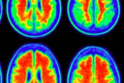
With the help of PET imaging, researchers at the University of Cambridge believe they have confirmed the process by which tau proteins spread in the brain to advance Alzheimer's disease, according to a study published on 5 January in the journal Brain.
The buildup of tau and another abnormal protein, beta amyloid, have been linked to the development of Alzheimer's disease. The theory is beta amyloid occurs first, which encourages the appearance and spread of tau, and destroys nerve cells thus leading to cognitive and memory problems.
One theory is tau develops first in one location in the brain and then spreads to another, with the hypothesis known as transneuronal spread. There are, however, other schools of thought that postulate tau is produced in nerve cells, or that a lack of nutrients to the brain region could be the cause. Thanks to the power of PET, researchers can now compare the viability of these different theories.
In this study, first author Dr. Thomas Cope and colleagues analyzed functional connections in the brains of 17 Alzheimer's patients to evaluate how they are wired using PET with a fluorine-18-AV-1451 radiopharmaceutical. The resulting PET images supported transneuronal spread as the culprit of dementia.
"If the idea of transneuronal spread is correct, then the areas of the brain that are most highly connected should have the largest buildup of tau and will pass it on to their connections," Cole said.
The findings suggest the progression of Alzheimer's disease may be slowed or stopped through drug therapy of tau as it travels along neurons.
To see if the same phenomenon occurred with other neurodegenerative diseases, the researchers looked at 17 patients afflicted with progressive supranuclear palsy (PSP), a rare condition that affects balance, speech, and vision, but not memory. With this disease, tau tends to collect only at the base of the brain rather than throughout the organ.
The researchers found tau buildup in the PSP patients supported the alternative hypotheses to tau distribution rather than the transneuronal spread theory. In another finding, the researchers found brain connections became more random with greater tau buildup, which could explain the confusion and muddled memories of Alzheimer's patients, the researchers reported.


![Examples of ultrasound findings and techniques. (A) Images in a 39-year-old male patient with a mass in the left thigh. The mass is heterogeneous on the B-mode US image (compared with the patient in D) and showed increased microvascularity (superb microvascular imaging [SMI]) and shear-wave elastography (SWE) values. Undifferentiated pleomorphic sarcoma was diagnosed at biopsy (with pleomorphic rhabdomyosarcoma in surgical specimen). (B) Images in an 18-year-old male patient with a mass in the left leg. The mass is hypoechoic on the B-mode image, with no other findings suggestive of malignancy. The lesion is in contact with the cortex of the tibia, which is slightly irregular. CT revealed a doubtful anteromedial tibial erosion. The microvascular study demonstrated high vascularization, suggestive of malignancy. Periosteal Ewing sarcoma was diagnosed with both histologic and immunohistochemical confirmation. (C) Images in a 69-year-old female patient with a lump growing on the outside of the left leg. Multiple SWE examinations were performed (please note the high values obtained in the measurements, whereas the color map highlights the stiffness relative to adjacent tissues). SMI showed areas of increased vascularization to target for sampling. Undifferentiated spindle cell sarcoma was diagnosed at biopsy, with residual leiomyosarcoma in the surgical specimen after neoadjuvant therapy. (D) Images in a 56-year-old female patient with a mass in the right thigh. The mass is heterogeneous at both B-mode ultrasound (similar to patient A) and MRI (coronal T2-weighted spectral attenuated inversion recovery [SPAIR]; T1-weighted pre-contrast and postcontrast imaging), which even shows uptake after the administration of paramagnetic contrast material, which is traditionally suggestive of malignancy. Low values at SMI and elastography are suggestive of benignity. Spindle cell lipoma was diagnosed at biopsy, with atypical spindle cell lipomatous tumor in the surgical specimen.](https://img.auntminnieeurope.com/mindful/smg/workspaces/default/uploads/2026/08/images-radiol250278fig2.APCFLSvX6p.jpg?auto=format%2Ccompress&fit=crop&h=100&q=70&w=100)


![Examples of ultrasound findings and techniques. (A) Images in a 39-year-old male patient with a mass in the left thigh. The mass is heterogeneous on the B-mode US image (compared with the patient in D) and showed increased microvascularity (superb microvascular imaging [SMI]) and shear-wave elastography (SWE) values. Undifferentiated pleomorphic sarcoma was diagnosed at biopsy (with pleomorphic rhabdomyosarcoma in surgical specimen). (B) Images in an 18-year-old male patient with a mass in the left leg. The mass is hypoechoic on the B-mode image, with no other findings suggestive of malignancy. The lesion is in contact with the cortex of the tibia, which is slightly irregular. CT revealed a doubtful anteromedial tibial erosion. The microvascular study demonstrated high vascularization, suggestive of malignancy. Periosteal Ewing sarcoma was diagnosed with both histologic and immunohistochemical confirmation. (C) Images in a 69-year-old female patient with a lump growing on the outside of the left leg. Multiple SWE examinations were performed (please note the high values obtained in the measurements, whereas the color map highlights the stiffness relative to adjacent tissues). SMI showed areas of increased vascularization to target for sampling. Undifferentiated spindle cell sarcoma was diagnosed at biopsy, with residual leiomyosarcoma in the surgical specimen after neoadjuvant therapy. (D) Images in a 56-year-old female patient with a mass in the right thigh. The mass is heterogeneous at both B-mode ultrasound (similar to patient A) and MRI (coronal T2-weighted spectral attenuated inversion recovery [SPAIR]; T1-weighted pre-contrast and postcontrast imaging), which even shows uptake after the administration of paramagnetic contrast material, which is traditionally suggestive of malignancy. Low values at SMI and elastography are suggestive of benignity. Spindle cell lipoma was diagnosed at biopsy, with atypical spindle cell lipomatous tumor in the surgical specimen.](https://img.auntminnieeurope.com/mindful/smg/workspaces/default/uploads/2026/08/images-radiol250278fig2.APCFLSvX6p.jpg?auto=format%2Ccompress&fit=crop&h=112&q=70&w=112)