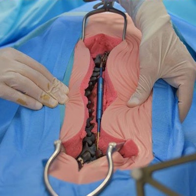
Surgeons in France used a 3D-printed model of the spine to simulate and hone a new robot-guided procedure on a 6-year-old boy with severe scoliosis. The young patient had such a severe combination of progressive scoliosis and spinal muscular atrophy (a neuromuscular disorder) that he was unable even to sit down.
Due to the complexity of the child's condition, a team of surgeons from Amiens-Picardie University Hospital spent more than a year preparing a specialized operation that employed a new robot-guided technique. Pediatric orthopedic surgeon Dr. François Deroussen, neurosurgeon Dr. Michel Lefranc, and head of the hospital's pediatric surgery department Dr. Richard Gouron, PhD, led the planning process.
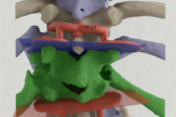
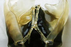
The 3D-printed model of the spine. All image courtesy of Amiens-Picardie University Hospital.A major component of the preparation included practicing the new surgery, which the physicians accomplished using 3D printing. First, the group created a 3D reconstruction of the child's chest cavity, and then they used that information to print an exact replica of the patient's spine at the SimUSanté training center affiliated with the hospital. Finally, they situated the resulting 3D-printed spine model into a mannequin the same size as the child.
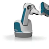
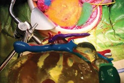
X-ray displaying spinal stem implants.The team used their 3D-printed model of the child's spine to simulate a robot-guided minimally invasive procedure, which enabled them to perfect their timing and fine-tune their surgical precision. The challenge of inserting 7-mm iliosacral screws into a bone corridor that was 8-mm wide proved possible with the support of a robotic surgical assistant (ROSA, MedTech).
Simulating the surgery on a 3D-printed model ultimately decreased actual operation time and minimized the risk of complications, according to the group. The success of the procedure helped prevent the onset of potential complications of scoliosis and also allowed the child to sit comfortably for the first time. The actual surgery took place on 28 September.
Amiens-Picardie University Hospital has already scheduled four other young patients for similar procedures. Moreover, the team of surgeons is working on formalizing their protocol so that other institutions can begin training in these techniques.


![Examples of ultrasound findings and techniques. (A) Images in a 39-year-old male patient with a mass in the left thigh. The mass is heterogeneous on the B-mode US image (compared with the patient in D) and showed increased microvascularity (superb microvascular imaging [SMI]) and shear-wave elastography (SWE) values. Undifferentiated pleomorphic sarcoma was diagnosed at biopsy (with pleomorphic rhabdomyosarcoma in surgical specimen). (B) Images in an 18-year-old male patient with a mass in the left leg. The mass is hypoechoic on the B-mode image, with no other findings suggestive of malignancy. The lesion is in contact with the cortex of the tibia, which is slightly irregular. CT revealed a doubtful anteromedial tibial erosion. The microvascular study demonstrated high vascularization, suggestive of malignancy. Periosteal Ewing sarcoma was diagnosed with both histologic and immunohistochemical confirmation. (C) Images in a 69-year-old female patient with a lump growing on the outside of the left leg. Multiple SWE examinations were performed (please note the high values obtained in the measurements, whereas the color map highlights the stiffness relative to adjacent tissues). SMI showed areas of increased vascularization to target for sampling. Undifferentiated spindle cell sarcoma was diagnosed at biopsy, with residual leiomyosarcoma in the surgical specimen after neoadjuvant therapy. (D) Images in a 56-year-old female patient with a mass in the right thigh. The mass is heterogeneous at both B-mode ultrasound (similar to patient A) and MRI (coronal T2-weighted spectral attenuated inversion recovery [SPAIR]; T1-weighted pre-contrast and postcontrast imaging), which even shows uptake after the administration of paramagnetic contrast material, which is traditionally suggestive of malignancy. Low values at SMI and elastography are suggestive of benignity. Spindle cell lipoma was diagnosed at biopsy, with atypical spindle cell lipomatous tumor in the surgical specimen.](https://img.auntminnieeurope.com/mindful/smg/workspaces/default/uploads/2026/08/images-radiol250278fig2.APCFLSvX6p.jpg?auto=format%2Ccompress&fit=crop&h=100&q=70&w=100)


![Examples of ultrasound findings and techniques. (A) Images in a 39-year-old male patient with a mass in the left thigh. The mass is heterogeneous on the B-mode US image (compared with the patient in D) and showed increased microvascularity (superb microvascular imaging [SMI]) and shear-wave elastography (SWE) values. Undifferentiated pleomorphic sarcoma was diagnosed at biopsy (with pleomorphic rhabdomyosarcoma in surgical specimen). (B) Images in an 18-year-old male patient with a mass in the left leg. The mass is hypoechoic on the B-mode image, with no other findings suggestive of malignancy. The lesion is in contact with the cortex of the tibia, which is slightly irregular. CT revealed a doubtful anteromedial tibial erosion. The microvascular study demonstrated high vascularization, suggestive of malignancy. Periosteal Ewing sarcoma was diagnosed with both histologic and immunohistochemical confirmation. (C) Images in a 69-year-old female patient with a lump growing on the outside of the left leg. Multiple SWE examinations were performed (please note the high values obtained in the measurements, whereas the color map highlights the stiffness relative to adjacent tissues). SMI showed areas of increased vascularization to target for sampling. Undifferentiated spindle cell sarcoma was diagnosed at biopsy, with residual leiomyosarcoma in the surgical specimen after neoadjuvant therapy. (D) Images in a 56-year-old female patient with a mass in the right thigh. The mass is heterogeneous at both B-mode ultrasound (similar to patient A) and MRI (coronal T2-weighted spectral attenuated inversion recovery [SPAIR]; T1-weighted pre-contrast and postcontrast imaging), which even shows uptake after the administration of paramagnetic contrast material, which is traditionally suggestive of malignancy. Low values at SMI and elastography are suggestive of benignity. Spindle cell lipoma was diagnosed at biopsy, with atypical spindle cell lipomatous tumor in the surgical specimen.](https://img.auntminnieeurope.com/mindful/smg/workspaces/default/uploads/2026/08/images-radiol250278fig2.APCFLSvX6p.jpg?auto=format%2Ccompress&fit=crop&h=112&q=70&w=112)