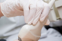
Using ultrasound to evaluate changes to the skeletal muscles after trauma may help physicians predict how patients will do once they have been released from the intensive care unit (ICU), according to an Italian study published online on 6 October in Annals of Intensive Care.
This kind of ultrasound data could also help clinicians determine how to prevent loss of muscle mass and function in these patients, wrote the group led by Dr. Maria Annetta of Gemelli University Hospital in Rome.
"We were able to quantify the morphological changes of skeletal muscle in trauma patients," the authors wrote. "Further studies may rely on this technique to evaluate the impact of different therapeutic strategies on muscle wasting."
Early intervention
Loss of muscle mass and function after traumatic injury is associated with worse short- and long-term outcomes. It starts within the first 24 hours after admission to intensive care and can continue for years, Annetta and colleagues wrote. Long-term muscle impairment may contribute to physical, mental, and cognitive dysfunction, affecting the quality of life of trauma survivors and also boosting healthcare costs.
 Dr. Maria Annetta of Gemelli University Hospital in Rome.
"Early physical rehabilitation has been associated with conflicting results in terms of functional outcome, so that the best strategy would theoretically be to avoid or minimize muscle loss during ICU stay," the group wrote. "B-mode ultrasonographic evaluation of skeletal muscles ... is an emerging and reliable tool to assess muscle changes over time."
The study included 38 severely injured patients admitted to the intensive care unit after trauma who stayed for up to three weeks. Patients' injuries were assessed according to the Injury Severity Score (ISS), which standardizes the severity of traumatic injury based on the worst injury of six body systems, based on a scale from 0 to 75. The median ISS score for the study cohort was 34.
The researchers focused specifically on ultrasound assessment of the rectus femoris and anterior tibialis muscles in young patients (median age, 40). The patients were tube fed as soon as they were stable and fully resuscitated, usually within 24 hours, the group wrote; some patients received a standard feeding formula, while others received a high-protein formula. Patients underwent ultrasound evaluation within 24 hours of the trauma event (day 0) and again at five days, 10 days, 15 days, and 20 days.
To measure muscle changes, the researchers tracked the anterior-posterior and lateral-lateral diameters of the muscles, as well as their cross-sectional area, which is calculated from the perimetral contour of the muscle area and is considered to be proportional to the total mass of the skeletal muscle.
Ultrasound evaluation showed that rectus femoris muscle mass changed significantly over the course of patients' ICU stay, with anterior-posterior diameter decreasing progressively between day 5 and day 20. Lateral-lateral diameter did not show a progressive decrease, but the difference between day 0 and day 20 was significant, the group noted. The cross-sectional area of the rectus femoris muscle progressively decreased as well, particularly between day 5 and day 20, with an overall loss of cross-sectional area of 45%.
As for anterior tibialis muscles, the anterior-posterior diameter decreased progressively over the course of the ICU stay for all patients, although lateral-lateral diameter did not decrease significantly. Overall loss of cross-sectional area for the anterior tibialis muscles was 22%; however, this was not statistically significant, according to the researchers.
In addition, there was a progressive increase in both rectus femoris and anterior tibialis echogenicity from day 0 onward, Annetta and colleagues found. An increase in echogenicity is associated with muscle cell depletion. The researchers did not find significant muscle mass differences in patients according to feeding formula, which led them to hypothesize that muscle loss may be caused by immobilization and inflammation rather than inadequate nutritional support.
"Inactivity is a potent stimulus to muscle protein breakdown," the group wrote. "Immobility of limbs is quite common in ICU patients and is related to bed rest and sedation. Acute and chronic activation of [the] inflammatory pathway is another potent stimulus for [this protein breakdown]."
The study shows that ultrasound is an effective tool for assessing muscle changes in ICU patients, although how to minimize loss of muscle mass in ICU patients needs further research, Annetta told AuntMinnie.com via email.
"Our research demonstrates that ultrasound can be used to monitor the quantitative and qualitative muscle changes over time in ICU patients -- and to assess whether interventions such as nutrition, physical therapy, or anabolic agents have any impact on muscle mass and structure," she said.


