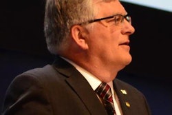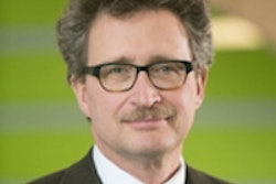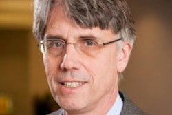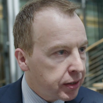
Dr. Stefan Schönberg became president of the German Radiological Society (Deutsche Röntgengesellschaft, DRG) earlier this year. He has been director of the Institute for Clinical Radiology and Nuclear Medicine at University Hospital Mannheim since 2007, and his main research interests are vascular and abdominal imaging, functional and high-field MRI, and oncologic imaging.
In this interview, Schönberg reflects on the changes taking place in the fast-moving world of medical imaging and shares his thoughts on the challenges ahead.
What plans do you have for your term as president?
 Dr. Stefan Schönberg from Mannheim.
Schönberg: The great development happening today, and which will be my principal focus during my term, is that whereas in Röntgen's time there was an image and a screen, behind the ray there is now a detector, a sensor, with many tiny packets of data flowing from this sensor; and that means so much data are produced that we can no longer fully utilize them all.
As the human brain can't process them all, we now have machines to help us with this.
In Röntgen's day, all you could see was a bone fracture on a radiograph, whereas in the future we will be able to use these data to predict how quickly a fracture will heal, whether a cancer patient will benefit from a new drug, and perhaps also how long they can expect to live.
What's become of the x-ray since 1895?
We have dramatically reduced the radiation dose. We have learned how to use all the properties of these rays even better. We can now use a range of energies to produce detailed imaging of tissues and we have also learned how to combine x-rays with other procedures and other great discoveries, such as MRI.
So is this procedure still regarded with suspicion?
I think we've learned a lot from that and x-rays are now widely accepted. Many people know that the only way we can achieve a precise diagnosis for the patient is to use these x-rays, but, as I said, we've also done a lot of research into side-effects and can now achieve a high degree of precision. However -- and this reflects our society today -- there is a great need for safety, which we can also take into account.
How does radiology contribute to the development of medicine?
Continuous improvements in diagnosis and more precise diagnostic investigations and minimally invasive therapy, particularly in cancers and also in vascular disease, have given radiology an increasing presence throughout almost the entire process of diagnosis and treatment. And, of course, a particular strength is that with the digital transformation, all areas of medicine now have a special opportunity to tailor this diagnosis and treatment to the individual patient.
What special challenges does this bring? With regard to tumor heterogeneity, for instance?
This is the thing: We know tumors can also pass on their genetic information, for instance when they spread within the body or form metastases. The big problem is the genetic material of one metastasis can be quite different from that of another, and this often makes medicines less than 100% effective.
In radiology we can use existing procedures, but refined and improved, in combination with modern information technology to determine whether a tumor will or will not respond to medication. If it does not do so, we can still act supportively, with our minimally invasive treatments.
What do you think should be the role of the DRG in this context?
The DRG is devoting an enormous amount of attention to this topic and we are now building up a large research platform to systematically extract precisely this knowledge from the data and to implement learning technologies, for instance, to deliver these procedures more widely.
Do you have a personal message for your colleagues?
The radiology community will have a special role, e.g., to help shape precision medicine, or medicine tailored to the patient, using these more refined diagnostic procedures and more precise treatment methods.
And how will things evolve beyond that? In 150 years, say?
It's difficult to predict that far ahead, but I believe the spirit of innovation must continue unbroken.
Editor's note: This is an edited version of a translation of an interview published in German online by the German Radiological Society (DRG, Deutsche Röntgengesellschaft). Translation by Syntacta Translation & Interpreting. To read the interview in German, click here. Some extracts came from a video interview posted by the Healthcare IT Media Portal site. For the original version, click here.