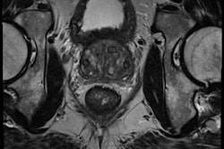
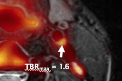
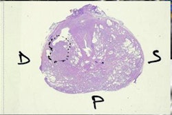
Thanks to recent research demonstrating its high sensitivity and negative predictive value for clinically significant prostate cancers, multiparametric MRI (MP-MRI) shows potential as a screening triage tool for identifying these all-important cases, according to a new editorial published in BMJ.
Prostate cancer screening is a controversial topic, as many cancers detected during screening have a protracted and indolent natural course. These patients are at greater risk from the ensuing cascade of diagnostic imaging studies and unnecessary treatments than from the disease itself, according to Dr. Philipp Dahm, a professor of surgery in the urology section at the University of Minnesota in the U.S. (BMJ, 19 September 2017).
However, MP-MRI has been shown to be highly sensitive in identifying aggressive cancers. Dahm noted the results from the Prostate MR Imaging Study (PROMIS), a U.K. trial that found that MP-MRI was almost twice as sensitive as transrectal ultrasound-guided (TRUS) biopsies at detecting clinically significant cancer. The modality also had a negative predictive value of 89%, and the researchers estimated that more than 25% of men could avoid biopsy based on MP-MRI findings.
While PROMIS provided only indirect evidence for how MRI would perform as a triage test in the diagnostic pathway, the same research group is now conducting a randomized controlled trial to evaluate the performance of triage MP-MRI, Dahm said. In the Prostate Evaluation for Clinically Important Disease: Sampling Use Image-Guidance or Not (PRECISION) trial, 470 men will be randomized to receive either standard 12-core TRUS biopsy or MP-MRI triage. Patients in the MP-MRI triage group will only be given a prostate biopsy after a suspicious MP-MRI, he noted. MP-MRI would then be used to guide the biopsy.
"Men at risk of prostate cancer, their families, and the health professionals who care for them should all be encouraged by the development of [MP-MRI] and the rigorous evidence-based approach being taken to evaluate new screening strategies," Dahm wrote. "While not solving all current problems, both hold much promise for the future of prostate cancer screening."


![Examples of ultrasound findings and techniques. (A) Images in a 39-year-old male patient with a mass in the left thigh. The mass is heterogeneous on the B-mode US image (compared with the patient in D) and showed increased microvascularity (superb microvascular imaging [SMI]) and shear-wave elastography (SWE) values. Undifferentiated pleomorphic sarcoma was diagnosed at biopsy (with pleomorphic rhabdomyosarcoma in surgical specimen). (B) Images in an 18-year-old male patient with a mass in the left leg. The mass is hypoechoic on the B-mode image, with no other findings suggestive of malignancy. The lesion is in contact with the cortex of the tibia, which is slightly irregular. CT revealed a doubtful anteromedial tibial erosion. The microvascular study demonstrated high vascularization, suggestive of malignancy. Periosteal Ewing sarcoma was diagnosed with both histologic and immunohistochemical confirmation. (C) Images in a 69-year-old female patient with a lump growing on the outside of the left leg. Multiple SWE examinations were performed (please note the high values obtained in the measurements, whereas the color map highlights the stiffness relative to adjacent tissues). SMI showed areas of increased vascularization to target for sampling. Undifferentiated spindle cell sarcoma was diagnosed at biopsy, with residual leiomyosarcoma in the surgical specimen after neoadjuvant therapy. (D) Images in a 56-year-old female patient with a mass in the right thigh. The mass is heterogeneous at both B-mode ultrasound (similar to patient A) and MRI (coronal T2-weighted spectral attenuated inversion recovery [SPAIR]; T1-weighted pre-contrast and postcontrast imaging), which even shows uptake after the administration of paramagnetic contrast material, which is traditionally suggestive of malignancy. Low values at SMI and elastography are suggestive of benignity. Spindle cell lipoma was diagnosed at biopsy, with atypical spindle cell lipomatous tumor in the surgical specimen.](https://img.auntminnieeurope.com/mindful/smg/workspaces/default/uploads/2026/08/images-radiol250278fig2.APCFLSvX6p.jpg?auto=format%2Ccompress&fit=crop&h=100&q=70&w=100)


![Examples of ultrasound findings and techniques. (A) Images in a 39-year-old male patient with a mass in the left thigh. The mass is heterogeneous on the B-mode US image (compared with the patient in D) and showed increased microvascularity (superb microvascular imaging [SMI]) and shear-wave elastography (SWE) values. Undifferentiated pleomorphic sarcoma was diagnosed at biopsy (with pleomorphic rhabdomyosarcoma in surgical specimen). (B) Images in an 18-year-old male patient with a mass in the left leg. The mass is hypoechoic on the B-mode image, with no other findings suggestive of malignancy. The lesion is in contact with the cortex of the tibia, which is slightly irregular. CT revealed a doubtful anteromedial tibial erosion. The microvascular study demonstrated high vascularization, suggestive of malignancy. Periosteal Ewing sarcoma was diagnosed with both histologic and immunohistochemical confirmation. (C) Images in a 69-year-old female patient with a lump growing on the outside of the left leg. Multiple SWE examinations were performed (please note the high values obtained in the measurements, whereas the color map highlights the stiffness relative to adjacent tissues). SMI showed areas of increased vascularization to target for sampling. Undifferentiated spindle cell sarcoma was diagnosed at biopsy, with residual leiomyosarcoma in the surgical specimen after neoadjuvant therapy. (D) Images in a 56-year-old female patient with a mass in the right thigh. The mass is heterogeneous at both B-mode ultrasound (similar to patient A) and MRI (coronal T2-weighted spectral attenuated inversion recovery [SPAIR]; T1-weighted pre-contrast and postcontrast imaging), which even shows uptake after the administration of paramagnetic contrast material, which is traditionally suggestive of malignancy. Low values at SMI and elastography are suggestive of benignity. Spindle cell lipoma was diagnosed at biopsy, with atypical spindle cell lipomatous tumor in the surgical specimen.](https://img.auntminnieeurope.com/mindful/smg/workspaces/default/uploads/2026/08/images-radiol250278fig2.APCFLSvX6p.jpg?auto=format%2Ccompress&fit=crop&h=112&q=70&w=112)