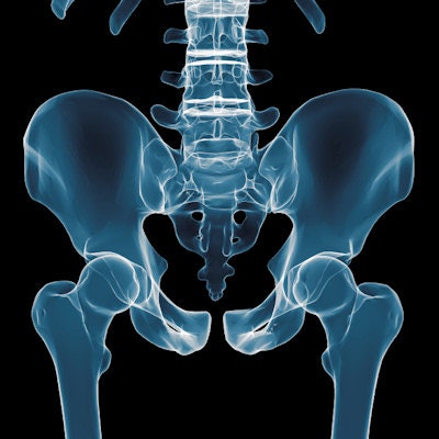
Researchers from Lebanon have found they can estimate the pelvic axial rotation of children and adults during x-ray acquisition, and any x-rays in which the axial rotation exceeds 10° should be discarded.
The group reconstructed x-rays obtained from lateral pelvic CT scans and simulated the axial rotation of the pelvis at various degrees to determine at what point the acceptable threshold of errors was reached. Ayman Assi, PhD, and colleagues from the Laboratory of Biomechanics and Medical Imaging and the University of Saint-Joseph in Beirut found the threshold is reached at 10° (Clinical Radiology, 22 April 2017).
"The reproducibility of sagittal pelvic parameters deteriorates when the axial rotation of the patient increases," they noted. "Biases on clinical parameters exceed the acceptable values when the axial rotation reaches 10°."
Why axial rotation?
The sagittal curvatures of the spine vary among asymptomatic subjects, and sagittal spine curvatures are highly correlated to pelvic morphological and positional parameters, according to the team. And pelvic parameters have become essential when assessing sagittal malalignment because of their strong relationship with spinal curvatures.
"For preoperative planning, the desired postoperative sagittal curvatures should be based on the morphology of the pelvis, quantified by pelvic incidence and sagittal pelvic thickness, as well as on other important positional parameters of the pelvis, namely sacral slope, pelvic tilt, and pelvic inclination," the study authors wrote.
These parameters are important in the radiographic evaluation of a range of spinal pathologies, such as spondylolisthesis, adolescent idiopathic scoliosis, and adult spinal deformity, as well as degenerative conditions, Assi and colleagues added.
The trouble is the measurements can be biased by a number of factors, including the 2D nature of the radiograph and the potential malpositioning of patients during radiograph acquisition, meaning it's crucial to quantify the errors associated with this technique. Axial rotation of the pelvis in the horizontal plane could influence the lateral projection of the pelvis and, thus, all the measured parameters.
"It would be useful for physicians to have a tool to easily estimate the axial rotation of patients during radiograph acquisition solely from the available lateral pelvic radiograph," they noted.
The group of researchers set out to do just that. They obtained lateral digitally reconstructed radiographs from the pelvic CT scans of eight children and nine adults, then simulated the axial rotation of the pelvis by rotating the CT volume around the vertical axis. They reconstructed the corresponding radiographs at 5°, 10°, 15°, and 20° of the axial rotation.
The researchers digitally measured the pelvic parameters on each radiograph and evaluated the intra- and interobserver variabilities at each axial rotation position. Three operators repeated the measurements three times each. The bias on each clinical parameter in each axial rotation position was calculated relative to the 0° position.
They found interobserver variability increased similarly in children and adults with axial rotation. It reached 4.4° for pelvic incidence and 4.7° for the sacral slope at 20° of axial rotation. Also, the higher the axial rotation, the more biases on radiological parameters. When axial rotation surpassed 10°, so did the acceptable threshold for errors.
The researchers also established a linear regression (R2 = 0.834, p < 0.0001) to estimate the axial rotation of a patient on a lateral pelvic radiograph based on the measurement of the bifemoral distance normalized to the sagittal pelvic thickness.
What this means
"Although it was expected that the bias on bifemoral distance increased with the axial rotation of the patient, the present study also reveals that the bias on the remaining parameters increased with this rotation, except for sagittal pelvic thickness, whose bias remained constant," the study authors wrote. "This can be explained by the fact that this parameter consists of the measurement of a line that is close to vertical, and, therefore, is not affected by the axial rotation of the patient."
No statistical differences existed on the biases between the children and adult groups, but it was noteworthy that the biases and their variances were consistently higher in the children's group. Sagittal pelvic parameters can be considered as reliable only if the axial rotation of the patient during radiograph acquisition was inferior to 10°, Assi and colleagues advised.
The equation the researchers formulated may be used in clinical practice as a tool for estimating axial rotation on any given lateral pelvic radiograph.
"Therefore, considering the previous results on the level of biases when axial rotation exceeds 10°, if the physician were to calculate an axial rotation equal or superior to this value, the radiograph should be repeated with correct patient positioning," they wrote.
The formula would also be applicable for any lateral radiograph as the distance is normalized and, thus, independent of the value of the enlargement factor, which occurs similarly in the vertical and horizontal directions.
The major limitation of the study is that the simulated radiographs were reconstructed from pelvic CT images. However, simulating the axial rotation circumvented the ethical constraint of exposing patients to multiple radiographs at different axial rotation positions, especially the children, the authors noted. Another limitation of the present study is the small number of subjects.


