Dear Cardiac Imaging Insider,
The European Society of Cardiology Congress (ESC 2016) held in Rome last week featured a trove of important studies that will inform our community now and in the weeks to come.
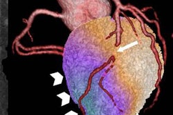
One recurring theme we noticed was research in hybrid cardiac imaging using two modalities, with the second test deployed to confirm the results of coronary CT angiography (CCTA) for suspected coronary artery disease. Considering the magnitude of the chest pain problem, and high associated costs of care, the focus seemed a good one.
One combination was the marriage of CCTA with SPECT myocardial perfusion imaging. Researchers from University Hospital Zurich in Switzerland looked at 400 chest pain patients who happened to undergo both tests, and they followed their progress for seven years. Matched findings on both exams, whether positive or negative, offered important prognostic information.
Another study presented at the meeting looked at how significantly costs and the number of invasive angiographies could be reduced with a CT-first approach. The six-nation, 18-center CONSERVE trial found large differences in everything but cardiac events. Get the rest of the story here.
Meanwhile, a massive Danish study presented at ESC 2016 enrolled almost 87,000 patients with chest pain, randomizing them evenly to an invasive or noninvasive diagnostic approach. The differences were stark and unequivocal.
Of course, some patients are tested before any hint of chest pain occurs and found to have atherosclerotic lesions outside the heart. Will treating extracoronary lesions aggressively confer better outcomes? Find out by clicking here.
Speaking of subtle, a recently developed cardiac MRI exam can find the earliest hint of left ventricular dysfunction, according to investigators from Sweden. Learn how it might be useful here. We also have a story about the use of femoral ultrasound in cardiovascular risk assessment. And while we're not on the subject, do you know how cardiologists use Twitter? You can find out here.
Last but not least, we invite you to scroll through the links below for the rest of the news from the heart of Europe, right here in your Cardiac Imaging Community.


![Examples of ultrasound findings and techniques. (A) Images in a 39-year-old male patient with a mass in the left thigh. The mass is heterogeneous on the B-mode US image (compared with the patient in D) and showed increased microvascularity (superb microvascular imaging [SMI]) and shear-wave elastography (SWE) values. Undifferentiated pleomorphic sarcoma was diagnosed at biopsy (with pleomorphic rhabdomyosarcoma in surgical specimen). (B) Images in an 18-year-old male patient with a mass in the left leg. The mass is hypoechoic on the B-mode image, with no other findings suggestive of malignancy. The lesion is in contact with the cortex of the tibia, which is slightly irregular. CT revealed a doubtful anteromedial tibial erosion. The microvascular study demonstrated high vascularization, suggestive of malignancy. Periosteal Ewing sarcoma was diagnosed with both histologic and immunohistochemical confirmation. (C) Images in a 69-year-old female patient with a lump growing on the outside of the left leg. Multiple SWE examinations were performed (please note the high values obtained in the measurements, whereas the color map highlights the stiffness relative to adjacent tissues). SMI showed areas of increased vascularization to target for sampling. Undifferentiated spindle cell sarcoma was diagnosed at biopsy, with residual leiomyosarcoma in the surgical specimen after neoadjuvant therapy. (D) Images in a 56-year-old female patient with a mass in the right thigh. The mass is heterogeneous at both B-mode ultrasound (similar to patient A) and MRI (coronal T2-weighted spectral attenuated inversion recovery [SPAIR]; T1-weighted pre-contrast and postcontrast imaging), which even shows uptake after the administration of paramagnetic contrast material, which is traditionally suggestive of malignancy. Low values at SMI and elastography are suggestive of benignity. Spindle cell lipoma was diagnosed at biopsy, with atypical spindle cell lipomatous tumor in the surgical specimen.](https://img.auntminnieeurope.com/mindful/smg/workspaces/default/uploads/2026/08/images-radiol250278fig2.APCFLSvX6p.jpg?auto=format%2Ccompress&fit=crop&h=100&q=70&w=100)


![Examples of ultrasound findings and techniques. (A) Images in a 39-year-old male patient with a mass in the left thigh. The mass is heterogeneous on the B-mode US image (compared with the patient in D) and showed increased microvascularity (superb microvascular imaging [SMI]) and shear-wave elastography (SWE) values. Undifferentiated pleomorphic sarcoma was diagnosed at biopsy (with pleomorphic rhabdomyosarcoma in surgical specimen). (B) Images in an 18-year-old male patient with a mass in the left leg. The mass is hypoechoic on the B-mode image, with no other findings suggestive of malignancy. The lesion is in contact with the cortex of the tibia, which is slightly irregular. CT revealed a doubtful anteromedial tibial erosion. The microvascular study demonstrated high vascularization, suggestive of malignancy. Periosteal Ewing sarcoma was diagnosed with both histologic and immunohistochemical confirmation. (C) Images in a 69-year-old female patient with a lump growing on the outside of the left leg. Multiple SWE examinations were performed (please note the high values obtained in the measurements, whereas the color map highlights the stiffness relative to adjacent tissues). SMI showed areas of increased vascularization to target for sampling. Undifferentiated spindle cell sarcoma was diagnosed at biopsy, with residual leiomyosarcoma in the surgical specimen after neoadjuvant therapy. (D) Images in a 56-year-old female patient with a mass in the right thigh. The mass is heterogeneous at both B-mode ultrasound (similar to patient A) and MRI (coronal T2-weighted spectral attenuated inversion recovery [SPAIR]; T1-weighted pre-contrast and postcontrast imaging), which even shows uptake after the administration of paramagnetic contrast material, which is traditionally suggestive of malignancy. Low values at SMI and elastography are suggestive of benignity. Spindle cell lipoma was diagnosed at biopsy, with atypical spindle cell lipomatous tumor in the surgical specimen.](https://img.auntminnieeurope.com/mindful/smg/workspaces/default/uploads/2026/08/images-radiol250278fig2.APCFLSvX6p.jpg?auto=format%2Ccompress&fit=crop&h=112&q=70&w=112)