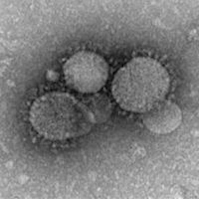
A single patient transmitted the Middle East respiratory syndrome coronavirus (MERS-CoV) to nearly half of those infected with the virus during the 2015 outbreak in South Korea, according to a study published online on July 8 in Lancet.
Researchers from Samsung Medical Center in Seoul retrospectively mapped the transmission of South Korea's first outbreak of MERS-CoV in 2015. They found that the "superspreader" patient was linked to 82 infections of other patients, visitors, and healthcare workers over a three-day period before he was isolated.
These cases represented 44% of the 186 total MERS cases during the outbreak and marked the highest transmission of a virus by a single person outside of the Middle East, according to the research team led by Dr. Sun Young Cho and Dr. Ji-Man Kang.
The 35-year-old patient had initially acquired MERS-CoV from the outbreak's index patient at another hospital. He was admitted to Samsung Medical Center on May 27, 2015, with no information on potential exposure to MERS-CoV, according to the researchers.
After the team reviewed closed-circuit video footage and electronic medical records, they determined that 1,576 people had been exposed to the patient in the emergency room (ER). Of these, 82 became infected with the virus: 33 patients, eight healthcare workers, and 41 visitors.
Patients in the same zone of the ER as the infected patient had the highest risk of infection. The following numbers of patients were infected within each group:
- Patients in the same zone of the ER: 23 (20%) of 117 were infected
- Visitors: 38 (6%) of 683 were infected
- Patients with brief exposure at the registration area or radiology suite of the ER: 3 (5%) of 58 were infected
- Healthcare workers: 5 (2%) of 218 were infected
- Patients who stayed in different zones of the ER: 4 (1%) of 500 were infected
An additional nine MERS-CoV cases were excluded from the analysis due to a lack of reliable data, according to the researchers.
The authors noted that as long as MERS-CoV transmission continues in the Middle East, a single traveler has the potential to cause similar outbreaks.
"Emergency preparedness and vigilance are crucial to the prevention of further large outbreaks in the future," they wrote. "Our report serves as an international alarm that preparedness in hospitals, laboratories, and governmental agencies is the key not only for MERS-CoV infections but also for other new emerging infectious diseases."


![Examples of ultrasound findings and techniques. (A) Images in a 39-year-old male patient with a mass in the left thigh. The mass is heterogeneous on the B-mode US image (compared with the patient in D) and showed increased microvascularity (superb microvascular imaging [SMI]) and shear-wave elastography (SWE) values. Undifferentiated pleomorphic sarcoma was diagnosed at biopsy (with pleomorphic rhabdomyosarcoma in surgical specimen). (B) Images in an 18-year-old male patient with a mass in the left leg. The mass is hypoechoic on the B-mode image, with no other findings suggestive of malignancy. The lesion is in contact with the cortex of the tibia, which is slightly irregular. CT revealed a doubtful anteromedial tibial erosion. The microvascular study demonstrated high vascularization, suggestive of malignancy. Periosteal Ewing sarcoma was diagnosed with both histologic and immunohistochemical confirmation. (C) Images in a 69-year-old female patient with a lump growing on the outside of the left leg. Multiple SWE examinations were performed (please note the high values obtained in the measurements, whereas the color map highlights the stiffness relative to adjacent tissues). SMI showed areas of increased vascularization to target for sampling. Undifferentiated spindle cell sarcoma was diagnosed at biopsy, with residual leiomyosarcoma in the surgical specimen after neoadjuvant therapy. (D) Images in a 56-year-old female patient with a mass in the right thigh. The mass is heterogeneous at both B-mode ultrasound (similar to patient A) and MRI (coronal T2-weighted spectral attenuated inversion recovery [SPAIR]; T1-weighted pre-contrast and postcontrast imaging), which even shows uptake after the administration of paramagnetic contrast material, which is traditionally suggestive of malignancy. Low values at SMI and elastography are suggestive of benignity. Spindle cell lipoma was diagnosed at biopsy, with atypical spindle cell lipomatous tumor in the surgical specimen.](https://img.auntminnieeurope.com/mindful/smg/workspaces/default/uploads/2026/08/images-radiol250278fig2.APCFLSvX6p.jpg?auto=format%2Ccompress&fit=crop&h=100&q=70&w=100)


![Examples of ultrasound findings and techniques. (A) Images in a 39-year-old male patient with a mass in the left thigh. The mass is heterogeneous on the B-mode US image (compared with the patient in D) and showed increased microvascularity (superb microvascular imaging [SMI]) and shear-wave elastography (SWE) values. Undifferentiated pleomorphic sarcoma was diagnosed at biopsy (with pleomorphic rhabdomyosarcoma in surgical specimen). (B) Images in an 18-year-old male patient with a mass in the left leg. The mass is hypoechoic on the B-mode image, with no other findings suggestive of malignancy. The lesion is in contact with the cortex of the tibia, which is slightly irregular. CT revealed a doubtful anteromedial tibial erosion. The microvascular study demonstrated high vascularization, suggestive of malignancy. Periosteal Ewing sarcoma was diagnosed with both histologic and immunohistochemical confirmation. (C) Images in a 69-year-old female patient with a lump growing on the outside of the left leg. Multiple SWE examinations were performed (please note the high values obtained in the measurements, whereas the color map highlights the stiffness relative to adjacent tissues). SMI showed areas of increased vascularization to target for sampling. Undifferentiated spindle cell sarcoma was diagnosed at biopsy, with residual leiomyosarcoma in the surgical specimen after neoadjuvant therapy. (D) Images in a 56-year-old female patient with a mass in the right thigh. The mass is heterogeneous at both B-mode ultrasound (similar to patient A) and MRI (coronal T2-weighted spectral attenuated inversion recovery [SPAIR]; T1-weighted pre-contrast and postcontrast imaging), which even shows uptake after the administration of paramagnetic contrast material, which is traditionally suggestive of malignancy. Low values at SMI and elastography are suggestive of benignity. Spindle cell lipoma was diagnosed at biopsy, with atypical spindle cell lipomatous tumor in the surgical specimen.](https://img.auntminnieeurope.com/mindful/smg/workspaces/default/uploads/2026/08/images-radiol250278fig2.APCFLSvX6p.jpg?auto=format%2Ccompress&fit=crop&h=112&q=70&w=112)