Unenhanced spectral imaging is an emerging x-ray technique that can measure tissue properties without requiring a contrast agent. Unlike standard x-ray imaging -- which cannot differentiate a thin, highly attenuating sample from a thicker, less attenuating material -- spectral imaging can make this distinction by considering both the actual attenuation and its energy dependence.
Spectral imaging could potentially be used to distinguish cysts from tumors during breast cancer screening, helping to address the relatively low specificity of x-ray mammography and reduce recalls. However, the introduction of spectral techniques for this purpose has been hindered by a lack of tissue x-ray attenuation data.
Collaboration from Sweden and the U.K. has developed a method to measure the energy-dependent x-ray attenuation of tissue using photon-counting spectral mammography. In a previous study, the researchers used this technique to measure the attenuation of cyst fluid. Now, they have applied the same approach to solid breast lesions, to establish whether the attenuation differs enough from that of cysts to distinguish the two lesion types (Physics in Medicine and Biology, 10 March 2016, Vol. 61:7, pp. 2595-2612).
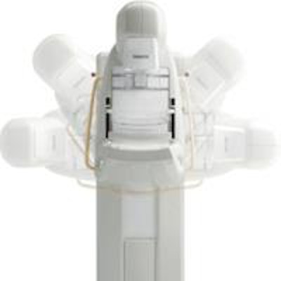
 Photograph and schematic of the Philips MicroDose SI mammography system.
"A spectral mammography system, the MicroDose SI, is already commercially available from Philips and is used in many clinics worldwide," said Erik Fredenberg, senior scientist at Philips Health Systems in Sweden. "The spectral data produced by the system are accessible to the advanced user, in addition to the spectral applications that are made available by Philips."
Attenuation measurements
Spectral mammography can be implemented by mapping a sample's attenuation to equivalent thicknesses of two reference materials -- in this study, aluminum (Al) and polymethyl methacrylate (PMMA). Fredenberg and co-investigators used the MicroDose SI to study five benign and 56 malignant solid breast lesions. During imaging, they positioned a step wedge made of Al and PMMA alongside the sample to provide a reference grid of thickness/material combinations. The sample's x-ray attenuation can then be determined by mapping the high- and low-energy counts obtained from a region-of-interest (ROI) against those obtained from ROIs on the step wedge.
The researchers acquired spectral images of ROIs in the samples and mapped the signals to equivalent thicknesses of the reference materials. They then plotted the Al-PMMA vector (equivalent PMMA thickness versus equivalent Al thickness) for all solid samples, as well as for previously published cyst fluid and water measurements.
 Equivalent PMMA and Al thickness normalized to 10 mm sample thickness, measured on 61 benign and malignant solid lesions, and compared with previously published data on cyst fluid and water. Left: Overview of the Al-PMMA vectors with the angle ï± and magnitude r indicated on the cyst-fluid vector. Right: Close-up of the measurement points. The five benign lesions are circled. For cyst fluid and water, the perimeters of the set of individual measurement points are shown.
No significant difference in attenuation was seen between benign and malignant solid breast lesions. However, the attenuation of solid breast lesions (benign and malignant) did differ significantly from that of cyst fluid, in terms of PMMA- and Al-equivalent thicknesses. Solid breast lesion attenuation also differed significantly from cyst attenuation in terms of the angle of the Al-PMMA vector, which is dependent upon the (effective) atomic number of the sample material, suggesting that the effective atomic number of cyst fluid is slightly higher than that of solid lesions.
The researchers also used the measured PMMA- and Al-equivalent thicknesses to calculate linear attenuation coefficients of the solid breast lesions, for a range of x-ray energies relevant to mammography. Comparing these with previously calculated linear attenuation coefficients for cyst fluid revealed almost overlapping data, with less than 2% difference on average in the 15 keV to 40 keV energy interval.
 Top: Linear attenuation coefficients of malignant solid breast lesions calculated from the measured PMMA- and Al-equivalent thicknesses, and compared with previously published data on cyst fluid and water. Bottom: The linear attenuation coefficients of malignant solid lesions and cyst fluid normalized to the linear attenuation coefficient of water.
However, they note it is still possible to distinguish between cyst fluid and solid lesions because the shapes of the linear attenuation curves are different (as clearly seen when data normalized to the linear attenuation coefficient of water are plotted).
Intra- and interimage variabilities did not differ significantly from the expected quantum noise, indicating that the sample homogeneity in each ROI and system stability were good. Compared with measurements on cyst fluid and water, the spread between different solid samples was relatively large, attributed mostly to natural variation of tumor tissue and only to a minor extent to random measurement errors and sample inhomogeneity.
The researchers concluded the significant separation between average cyst fluid and tumor attenuation suggests it may be possible to distinguish cystic from solid breast lesions using screening with spectral mammography. These results laid the groundwork for a pilot clinical study, the results of which were published earlier this year in Investigative Radiology.
"The results of the study were quite promising and we are starting up a large multisite trial, which will most likely be up and running before the end of 2016," Fredenberg told medicalphysicsweb.
© IOP Publishing Limited. Republished with permission from medicalphysicsweb, a community website covering fundamental research and emerging technologies in medical imaging and radiation therapy.