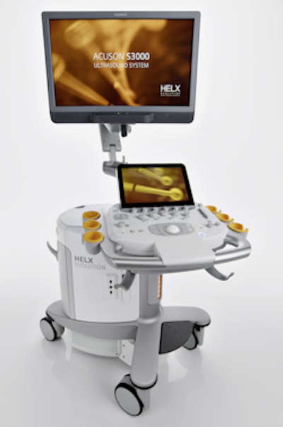
With the aim of improving the usability of high-end ultrasound, Siemens Healthcare has debuted the Helx Evolution with Touch Control upgrade for its Acuson S family of ultrasound systems at the Dreiländertreffen Ultraschall (DLT) conference in Davos, Switzerland.
In development since 2012, the hardware and software upgrade for the Acuson S1000, S2000, and S3000 scanners adds features such as a simplified user interface, a touchscreen display, improved probes, image quality enhancements, and more configuration options, according to the vendor. During the development process for the upgrade, the company visited 395 users around the world to learn from customers how it could best refine its ultrasound technology, said Douglas Younger, a product manager in Siemens' ultrasound business unit.
Helx Evolution with Touch Control upgrade as shown on an Acuson S3000 system. Image courtesy of Siemens Healthcare.Among the usability improvements is a simplified control panel that reduces the total number of tactile keys by about 33%. This change was achieved in part by combining different applications for a given function into a single button, according to the firm. As a result, users can directly select shear-wave elastography, 4D, and contrast-enhanced ultrasound studies.
The new 12-inch touchscreen is highly sensitive and can be used even while wearing latex gloves or with contact gel residue on the finger, Siemens said. The company has also added new configuration options to enable more flexible placement of body markers, for example, as well as a laser optical track ball to reduce repetitive stress injuries, dynamic backlighting, and an integrated, customizable gel warmer. An eSie Image feature offers real-time automatic image optimization, allowing users to focus on scanning, Siemens said.
In addition to workflow and usability enhancements, Helx Evolution with Touch Control also comes with a range of image quality improvements. The vendor's high-density ultrasound probes have been enhanced, featuring approximately double the crystals included in the standard probes available for the S family, Siemens said. This provides more-detailed images and, consequently, improvements in diagnostic accuracy, according to the vendor.
The company said it has also improved B-mode imaging capabilities, and enhanced ultrasound probe coordination allows the abdominal probe to provide deeper penetration. Rib shadowing has also been reduced, Siemens said.
An improvement to the vendor's high-frequency linear transducer has yielded better visualization for superficial scanning in breast imaging, for example, according to the company. Siemens said it has also improved cardiovascular imaging capabilities.
In addition, the new upgrade makes it easier to perform elastography applications available on the Acuson S line, Siemens said. B-mode images and elastography now appear side by side thanks to the upgrade, making it easier to identify pathological change on B-mode images, according to the firm.
Helx Evolution with Touch Control has received U.S. Food and Drug Administration (FDA) 510(k) clearance and the European CE Mark. Worldwide launch of the new systems is slated for later in the fall.
Siemens has not yet released pricing information.


![Examples of ultrasound findings and techniques. (A) Images in a 39-year-old male patient with a mass in the left thigh. The mass is heterogeneous on the B-mode US image (compared with the patient in D) and showed increased microvascularity (superb microvascular imaging [SMI]) and shear-wave elastography (SWE) values. Undifferentiated pleomorphic sarcoma was diagnosed at biopsy (with pleomorphic rhabdomyosarcoma in surgical specimen). (B) Images in an 18-year-old male patient with a mass in the left leg. The mass is hypoechoic on the B-mode image, with no other findings suggestive of malignancy. The lesion is in contact with the cortex of the tibia, which is slightly irregular. CT revealed a doubtful anteromedial tibial erosion. The microvascular study demonstrated high vascularization, suggestive of malignancy. Periosteal Ewing sarcoma was diagnosed with both histologic and immunohistochemical confirmation. (C) Images in a 69-year-old female patient with a lump growing on the outside of the left leg. Multiple SWE examinations were performed (please note the high values obtained in the measurements, whereas the color map highlights the stiffness relative to adjacent tissues). SMI showed areas of increased vascularization to target for sampling. Undifferentiated spindle cell sarcoma was diagnosed at biopsy, with residual leiomyosarcoma in the surgical specimen after neoadjuvant therapy. (D) Images in a 56-year-old female patient with a mass in the right thigh. The mass is heterogeneous at both B-mode ultrasound (similar to patient A) and MRI (coronal T2-weighted spectral attenuated inversion recovery [SPAIR]; T1-weighted pre-contrast and postcontrast imaging), which even shows uptake after the administration of paramagnetic contrast material, which is traditionally suggestive of malignancy. Low values at SMI and elastography are suggestive of benignity. Spindle cell lipoma was diagnosed at biopsy, with atypical spindle cell lipomatous tumor in the surgical specimen.](https://img.auntminnieeurope.com/mindful/smg/workspaces/default/uploads/2026/08/images-radiol250278fig2.APCFLSvX6p.jpg?auto=format%2Ccompress&fit=crop&h=100&q=70&w=100)


![Examples of ultrasound findings and techniques. (A) Images in a 39-year-old male patient with a mass in the left thigh. The mass is heterogeneous on the B-mode US image (compared with the patient in D) and showed increased microvascularity (superb microvascular imaging [SMI]) and shear-wave elastography (SWE) values. Undifferentiated pleomorphic sarcoma was diagnosed at biopsy (with pleomorphic rhabdomyosarcoma in surgical specimen). (B) Images in an 18-year-old male patient with a mass in the left leg. The mass is hypoechoic on the B-mode image, with no other findings suggestive of malignancy. The lesion is in contact with the cortex of the tibia, which is slightly irregular. CT revealed a doubtful anteromedial tibial erosion. The microvascular study demonstrated high vascularization, suggestive of malignancy. Periosteal Ewing sarcoma was diagnosed with both histologic and immunohistochemical confirmation. (C) Images in a 69-year-old female patient with a lump growing on the outside of the left leg. Multiple SWE examinations were performed (please note the high values obtained in the measurements, whereas the color map highlights the stiffness relative to adjacent tissues). SMI showed areas of increased vascularization to target for sampling. Undifferentiated spindle cell sarcoma was diagnosed at biopsy, with residual leiomyosarcoma in the surgical specimen after neoadjuvant therapy. (D) Images in a 56-year-old female patient with a mass in the right thigh. The mass is heterogeneous at both B-mode ultrasound (similar to patient A) and MRI (coronal T2-weighted spectral attenuated inversion recovery [SPAIR]; T1-weighted pre-contrast and postcontrast imaging), which even shows uptake after the administration of paramagnetic contrast material, which is traditionally suggestive of malignancy. Low values at SMI and elastography are suggestive of benignity. Spindle cell lipoma was diagnosed at biopsy, with atypical spindle cell lipomatous tumor in the surgical specimen.](https://img.auntminnieeurope.com/mindful/smg/workspaces/default/uploads/2026/08/images-radiol250278fig2.APCFLSvX6p.jpg?auto=format%2Ccompress&fit=crop&h=112&q=70&w=112)