New research indicates that PET imaging with the radiotracer florbetaben can detect a form of beta-amyloid plaque that develops early in the brain. The findings could possibly lead to the earlier detection of Alzheimer's disease, as well as new clinical indications for florbetaben.
In the study, which was presented earlier this month at the Society of Nuclear Medicine and Molecular Imaging (SNMMI) annual meeting in Baltimore, researchers found that PET with florbetaben (Neuraceq, Piramal Imaging) was able to detect diffuse beta-amyloid plaque in certain regions of the brain.
"This would support the fact that florbetaben can detect this initial plaque in the brain," lead author Dr. Ana Catafau, PhD, from the nuclear medicine department at Autonomous University of Barcelona and Hospital de Sant Pau in Spain, told AuntMinnie.com. "It also was very interesting to see that florbetaben can detect other types of plaques, besides neuritic plaques, which usually develop in the later stages of Alzheimer's."
Different types of plaque
The U.S. Food and Drug Administration (FDA) approved Neuraceq in 2014 for use with PET to assess neuritic beta-amyloid plaque in adult patients with cognitive impairment who are being evaluated for Alzheimer's disease and other causes of cognitive decline. The agent is not indicated for other types of deposits, such as diffuse and vascular beta-amyloid plaques.
But these types of plaque deposits may also be connected to Alzheimer's disease. Previous studies have suggested that diffuse beta-amyloid plaques are present in the early stages of Alzheimer's, while neuritic beta-amyloid plaque is more prevalent in the later stages of the disease.
Therefore, the researchers decided to examine the use of florbetaben for the different types of deposits. They collected brain tissue samples from 87 end-of-life patients, including 64 individuals with Alzheimer's disease, 14 people with other types of dementia, and nine elderly volunteers with no signs of dementia. The subjects had a mean age of 80.4 years (± 10.2 years) and had undergone florbetaben-PET scans before death.
"What we intended to do was to correlate the PET results in vivo with the brain autopsy after death," added Catafau, who is also vice president of clinical R&D for neurosciences at Piramal.
With those images, the researchers assessed neuritic, diffuse, and vascular beta-amyloid plaque in the frontal, occipital, anterior cingulate, and posterior cingulate cortices of the brain. Cortical standardized uptake value ratios (SUVRs) were obtained in all regions of interest using cerebellar gray matter as a reference region.
Florbetaben-PET discoveries
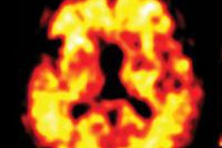
The PET scans revealed significant florbetaben uptake in the frontal and posterior cingulate cortices, with a high frequency of deposits of both diffuse and neuritic beta amyloid.
More importantly, in the occipital and anterior cingulate cortices, only diffuse beta-amyloid plaques contributed significantly to florbetaben uptake.
Meanwhile, the presence of vascular beta amyloid contributed significantly to florbetaben uptake only in the occipital region, the researchers found.
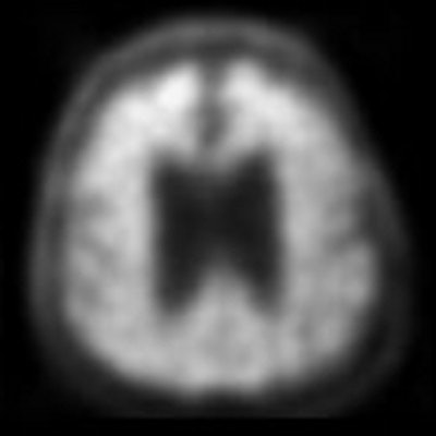
 Gray matter in the cerebellum was used as the reference region (top row). PET images of the frontal cortex (bottom row) show florbetaben uptake in diffuse and neuritic beta-amyloid plaques. Images courtesy of Dr. Ana Catafau, PhD.
"The message here is that with florbetaben we would be able to detect [Alzheimer's] disease at its early stages, even in cases where neuritic plaques may not be present," Catafau said.
The study's results could lead to an expansion of florbetaben's indications.
Andrew Stephens, chief medical officer at Piramal, noted that only 80% or so of people diagnosed with Alzheimer's disease have amyloid plaque.
"What this tells us is that the clinical diagnosis is not a perfect tool and the advantage of adding biomarkers can help improve diagnostic accuracy," he said.
It is "very important, as we move toward biomarkers for therapeutic compounds, that these earlier forms of plaques can be detected ... and may provide a more sensitive biomarker for the therapeutic trials looking at earlier stages of plaque," Stephens said.
Study disclosures
Piramal Imaging provided research support for the study.