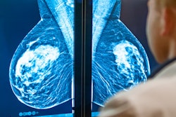
One-view digital breast tomosynthesis (DBT) detected 40% more breast cancers than traditional two-view mammography, with lower radiation dose, in a study of more than 7,500 women in Sweden that was published on 1 May in European Radiology.
In the Malmö Breast Tomosynthesis Screening Trial, researchers from Lund University compared one-view DBT with two-view full-field digital mammography (FFDM) that was independently double-read in a group of 7,500 individuals. The Malmö study is a population-based, prospective one-arm study that ultimately is planned to include 15,000 participants.
Lead author Dr. Kristina Lång notes that DBT has the potential to reduce the adverse effect of overlapping tissue on the interpretation of screening mammograms, and previous studies comparing two-view DBT with two-view digital mammography have shown that tomo has better performance. However, she and her colleagues wanted to examine whether DBT with a single mediolateral oblique (MLO) view would also be superior to digital mammography with both MLO and craniocaudal (CC) views.
A researcher team led by Dr. Sophia Zackrisson offered women ages 40-74 who were participating in the Swedish breast cancer screening program exams with single-view DBT in addition to conventional two-view digital mammography. Exams were performed with a commercially available DBT unit (Mammomat Inspiration, Siemens Healthcare).
Two-view digital mammography images followed by one-view DBT were acquired in a single screening session. The researchers estimated that the absorbed radiation dose of the DBT exam was 70% of that of the conventional mammography study. Six readers with experience in DBT image interpretation then read the exams, classifying findings on a five-point scale.
In all, breast cancer was found in 68 women, with both modalities detecting 46 cases. One-view DBT alone found 21 cases and digital mammography found one case alone. Other important characteristics are as follows:
| Digital mammography vs. one-view DBT per 1,000 screening exams | ||
| Two-view digital mammography | One-view DBT | |
| Cancer detection rate | 6.3 cancers | 8.9 cancers |
| Recall rate | 2.6% | 3.8% |
| Positive predictive value (PPV) | 24% | 24% |
The researchers concluded that their study indicated DBT was suitable for use as a screening exam that could replace conventional digital mammography. Also, cancers detected with DBT tended to be of lower grade, smaller size, and had not yet spread to the lymph nodes, and women were slightly younger at diagnosis. Another advantage was less breast compression was used with the DBT technique.
They noted their study was not designed to assess the differences between the modalities in terms of mortality, and that a very large, randomized study would be needed to prove any advantage. However, it could be possible to design a study that used a relative reduction of interval cancers as an end-point as a surrogate for screening efficacy.
"The main findings of this explorative analysis suggest that one-view DBT may be feasible as a standalone technique for breast cancer screening," the authors concluded. "Compared with the current gold standard (DM), DBT was superior in terms of detection rate and equal in terms of PPV."
In terms of future research, Lang said in an email to AuntMinnieEurope.com "Our aim is to understand the role of DBT and in particular one-view DBT as a standalone screening modality. In order to do that, we are trying to find some answers on the benefits and harms by studying several aspects such as false positives, interval cancers, trying to address the problem of overdiagnosis by studying tumor biology of the additional cancers detected, and last but not least, a cost-benefit analysis. We are inspired by the exciting results and are looking forward to the research that lies ahead."