Members of the Society of Radiographers (SoR) have announced their plans for a third strike to take place on 29 January from 8 a.m. to 2 p.m., followed by a work-to-rule period beginning on 30 January and continuing to 24 February.
There was no strike action in December as the unions decided not to place additional strain on the National Health Service (NHS) at this time, the society said. Because the government did not make a pay offer or attempt to meet with the NHS staff for negotiations, the unions have decided to strike in January.
The proposed strike and industrial action represents an escalation of previous actions with a six-hour strike this time and a longer period of work-to-rule. Other unions will be carrying out action in accordance with the mandates they have from their members, and some will be striking for 12 hours on 29 January.
SoR decided on six hours as the appropriate period that demonstrates an escalation of the dispute but will also enable members in both diagnostic imaging and radiotherapy to ensure, where possible, that while patients may be inconvenienced, they will not be put at risk, the society said.
If there is no change, unions will strike again on 25 February.
The society also plans to issue detailed guidance regarding emergency cover early next month to allow members and their managers to plan for this further strike.
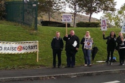
The proposed strike represents the third time the society will strike; the first was on 20 October and the second on 25 November.


![Overview of the study design. (A) The fully automated deep learning framework was developed to estimate body composition (BC) (defined as subcutaneous adipose tissue [SAT] in liters; visceral adipose tissue [VAT] in liters; skeletal muscle [SM] in liters; SM fat fraction [SMFF] as a percentage; and intramuscular adipose tissue [IMAT] in deciliters) from MRI. The fully automated framework comprised one model (model 1) to quantify different BC measures (SAT, VAT, SM, SMFF, and IMAT) as three-dimensional (3D) measures from whole-body MRI scans. The second model (model 2) was trained to identify standardized anatomic landmarks along the craniocaudal body axis (z coordinate field), which allowed for subdividing the whole-body measures into different subregions typically examined on clinical routine MRI scans (chest, abdomen, and pelvis). (B) BC was quantified from whole-body MRI in over 66,000 individuals from two large population-based cohort studies, the UK Biobank (UKB) (36,317 individuals) and the German National Cohort (NAKO) (30,291 individuals). Bar graphs show age distribution by sex and cohort. BMI = body mass index. (C) After the performance assessment of the fully automated framework, the change in BC measures, distributions, and profiles across age decades were investigated. Age-, sex-, and height-adjusted body composition reference curves were calculated and made publicly available in a web-based z-score calculator (https://circ-ml.github.io).](https://img.auntminnieeurope.com/mindful/smg/workspaces/default/uploads/2026/05/body-comp.XgAjTfPj1W.jpg?auto=format%2Ccompress&fit=crop&h=112&q=70&w=112)


