The development of postmortem CT angiography (PMCTA) represents a significant step forward to minimally invasive autopsies, but considerable work still has to be done before it can replace the conventional autopsy, Swiss researchers have found.
To help propel PMCTA from the research lab to routine practice, techniques need to be further investigated and standardized, and their artifacts, strengths, and pitfalls must be known and guidelines for the interpretation of the radiological data should be available. Only then can the techniques be used in sensitive medicolegal cases and can they be introduced into the workflow of a daily routine, noted Dr. Silke Grabherr, PhD, and colleagues, from the University Center of Legal Medicine Lausanne-Geneva, University Hospital of Lausanne in Switzerland.
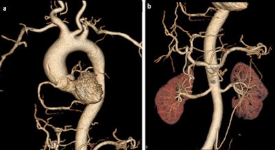 The quality of postmortem angiography using the technique of multiphase PMCTA is high. The vessels of the thorax (a) and abdomen (b) are visible in a volume-rendered 3D reconstruction. All images courtesy of Dr. Silke Grabherr, PhD.
An international working group, the Technical Working Group Post-mortem Angiography Methods (TWGPAM), was created in February 2012 to develop systematic and target-oriented research. It now consists of nine participating centers from six European countries: Switzerland (University of Lausanne and University of Basel), Germany (University of Hamburg, University of Munich, and University of Leipzig), the U.K. (University of Leicester), Italy (University of Foggia), Poland (University of Krakow), and France (University of Toulouse).
"Each center provides a team consisting of forensic pathologists, radiologists, and radiographers. The teams or their leaders meet regularly for meetings, are exchanging data with each other and report cases on a secured home page," the Swiss researchers wrote in an article posted online on 14 November by the British Journal of Radiology. "As the first research objective, the TWGPAM group has started a multicenter study in order to validate the technique of PMCTA. Each center should investigate about 50 cases using the standardized protocol of this method as well as standardized equipment (Virtangio perfusion device and consumables including the contrast agent Angiofil provided one year free of charge by Fumedica, Switzerland)."
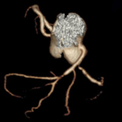
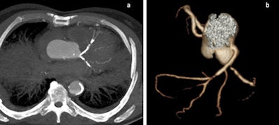 Coronary calcifications and stenoses are shown in 2D and 3D images in two cases of sudden cardiac death due to coronary artery disease.
One solution for implementing PMCTA into the daily routine of legal medicine is the introduction of radiographers to help with the additional examination. In fact, the forensic radiographer has become a new subspecialty in some countries, and international training is available, they noted.
"Beside the validation of the existing techniques, there are also further things to develop: Protocols for the investigation of superior and inferior members should be established, perfusion protocols for infants are needed, and methods for selective angiography other than the coronary arteries would be of interest. All these missing points underline that the research of PMCTA is still in the beginning," stated Grabherr et al.
The research group in Lausanne is doing multiple studies involving the technique of multiphase PMCTA (MPMCTA). Also, different research projects of the TWGPAM members are ongoing, such as studies about vascular lesions due to road traffic accidents, medicolegal investigations of medical errors using MPMCTA, and the use of MPMCTA for investigating cases of sharp trauma, as well as technical studies to investigate new selective cannulation approaches, etc.
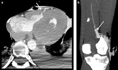 Reconstruction of sharp trauma lesions resulting from stab wounds. The first case (a) shows a stab wound trajectory in the chest with an intramyocardic trajectory. The second case (b) shows a stab wound in the left leg with a lesion of the popliteal artery (arrow in b).
The most important study at the moment is the validation study of MPMCTA, in which 500 cases investigated in different centers are being investigated. This study will be submitted for publication in spring 2014, Grabherr noted in an email to AuntMinnieEurope.com.
A large workshop about the technique of MPMCTA is planned for Hamburg, Germany, on 7-8 September 2014. Attendees will receive a lot of information about how to do the technique and what details can be derived from the images, and it will have a special focus on cases after cardiovascular and surgical intervention. It will be especially informative for cardiologists, radiologists, surgeons, pathologists, forensic pathologists, etc. There will be specialists from all different medical fields.
Study disclosures
The postmortem angiography project in the University Center of Legal Medicine Lausanne-Geneva was financially supported by the Promotion Agency for Innovation of the Swiss Confederation (KTI Nr.10221.1 PFIW-IW) from April 2009 to June 2011; Silke Grabherr has had personal academic funding from the Fondation Leenaards, Lausanne, Switzerland since April 2011; and the enterprise Fumedica, Muri, Switzerland, has provided consumables formultiphase postmortem CT angiography to the University Center of Legal Medicine Lausanne-Geneva in the context of an ongoing multicenter study started in February 2012.


