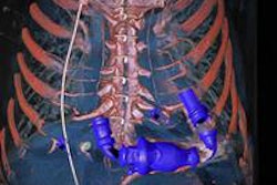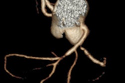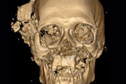
A team from the U.K. has reported that adding cardiac CT angiography (CCTA) to routine CT for postmortem imaging in suspected cases of death by coronary artery disease significantly increases the accuracy of virtual autopsies.
In a presentation at the recent U.S. and Canadian Academy of Pathology's 2013 annual meeting, Dr. Ian S. D. Roberts, a professor of cellular pathology at the John Radcliffe Hospital in Oxford, showed full-body CT coupled with an external examination followed by CCTA led to a firm diagnosis of ischemic heart disease as the cause of death in 23 individuals. In contrast, CT without angiography diagnosed only three of the 28 cases of coronary artery disease.
"With coronary angiography we have overcome the most important weakness of [postmortem] imaging," he told AuntMinnieEurope.com. "Another one remains: the diagnosis of pulmonary embolism. We have developed a method of pulmonary angiography and are validating it at the moment. The critical issue is being able to identify which cases you can correctly diagnose on imaging without autopsy -- we now have the experience to do that."
Roberts was the lead investigator in a study published last year in the Lancet that showed postmortem CT is more accurate than MRI in determining the cause of death, and that the error rate in radiologists' determination of cause of death is similar to that for clinical death certificates (January 2012, Vol. 379:9111, pp. 136-142). However, CT missed 100% (10/10) of the cases of fatal pulmonary embolism and 14% (12/86) of the cases of coronary artery disease as the cause of death.
The Oxford group developed a technique to use postmortem CCTA to determine whether coronary artery disease is the cause of death (Clinical Radiology, July 2011, Vol. 66:7, pp. 645-65). Along with Dr. Zoe Traill, Roberts then retrospectively compared the results of 60 coroner's cases examined with CT alone with another 60 cases examined with CT plus CCTA, from the period of February 2011 to August 2012. The CCTA was added when CT did not provide a definitive cause of death. The researchers based their definitive imaging determination of the cause of death on their experience with 300 previous cases in which they had correlated the results of postmortem CT and autopsy.
They found 62% of the cases imaged by CT alone required an autopsy to confirm the cause of death, compared with only 30% of cases imaged by CT plus CCTA. Coronary artery disease was the most common cause of death in both sets of cases, at 20 (33%) and 28 (47%), respectively.
However, CT alone resulted in only three of these cases being uncovered, two as coronary artery disease and another two as hemopericardium due to ruptured myocardial infarction, while CT plus CCTA uncovered 23 of the 28.
"This doesn't mean the other five cases were missed [by CT plus CCTA]," Roberts noted. "The coronary lesions were seen on angiography but there were other findings that required autopsy to investigate, or the clinical history suggested another potential cause of death."
Overall, they made a definite diagnosis based on imaging with CT alone in 38% of cases, and with CT plus CCTA in 70% of cases. For CT alone, there were 13 cases in which there was a major discrepancy between death diagnosis on imaging and autopsy, while there were eight with CT plus CCTA.
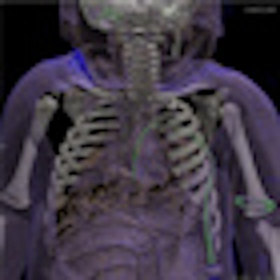
Editor's Note: Postmortem CT image on home page is of a 4-year-old child who was revived at the emergency department. An artery tube was left open after the procedure and the arteries were filled with air, but the child died on the hospital ward. Image courtesy of Dr. Anders Persson, PhD, Center for Medical Image Science and Visualization, Linköpings Universitet, Sweden.


![Examples of ultrasound findings and techniques. (A) Images in a 39-year-old male patient with a mass in the left thigh. The mass is heterogeneous on the B-mode US image (compared with the patient in D) and showed increased microvascularity (superb microvascular imaging [SMI]) and shear-wave elastography (SWE) values. Undifferentiated pleomorphic sarcoma was diagnosed at biopsy (with pleomorphic rhabdomyosarcoma in surgical specimen). (B) Images in an 18-year-old male patient with a mass in the left leg. The mass is hypoechoic on the B-mode image, with no other findings suggestive of malignancy. The lesion is in contact with the cortex of the tibia, which is slightly irregular. CT revealed a doubtful anteromedial tibial erosion. The microvascular study demonstrated high vascularization, suggestive of malignancy. Periosteal Ewing sarcoma was diagnosed with both histologic and immunohistochemical confirmation. (C) Images in a 69-year-old female patient with a lump growing on the outside of the left leg. Multiple SWE examinations were performed (please note the high values obtained in the measurements, whereas the color map highlights the stiffness relative to adjacent tissues). SMI showed areas of increased vascularization to target for sampling. Undifferentiated spindle cell sarcoma was diagnosed at biopsy, with residual leiomyosarcoma in the surgical specimen after neoadjuvant therapy. (D) Images in a 56-year-old female patient with a mass in the right thigh. The mass is heterogeneous at both B-mode ultrasound (similar to patient A) and MRI (coronal T2-weighted spectral attenuated inversion recovery [SPAIR]; T1-weighted pre-contrast and postcontrast imaging), which even shows uptake after the administration of paramagnetic contrast material, which is traditionally suggestive of malignancy. Low values at SMI and elastography are suggestive of benignity. Spindle cell lipoma was diagnosed at biopsy, with atypical spindle cell lipomatous tumor in the surgical specimen.](https://img.auntminnieeurope.com/mindful/smg/workspaces/default/uploads/2026/08/images-radiol250278fig2.APCFLSvX6p.jpg?auto=format%2Ccompress&fit=crop&h=100&q=70&w=100)


![Examples of ultrasound findings and techniques. (A) Images in a 39-year-old male patient with a mass in the left thigh. The mass is heterogeneous on the B-mode US image (compared with the patient in D) and showed increased microvascularity (superb microvascular imaging [SMI]) and shear-wave elastography (SWE) values. Undifferentiated pleomorphic sarcoma was diagnosed at biopsy (with pleomorphic rhabdomyosarcoma in surgical specimen). (B) Images in an 18-year-old male patient with a mass in the left leg. The mass is hypoechoic on the B-mode image, with no other findings suggestive of malignancy. The lesion is in contact with the cortex of the tibia, which is slightly irregular. CT revealed a doubtful anteromedial tibial erosion. The microvascular study demonstrated high vascularization, suggestive of malignancy. Periosteal Ewing sarcoma was diagnosed with both histologic and immunohistochemical confirmation. (C) Images in a 69-year-old female patient with a lump growing on the outside of the left leg. Multiple SWE examinations were performed (please note the high values obtained in the measurements, whereas the color map highlights the stiffness relative to adjacent tissues). SMI showed areas of increased vascularization to target for sampling. Undifferentiated spindle cell sarcoma was diagnosed at biopsy, with residual leiomyosarcoma in the surgical specimen after neoadjuvant therapy. (D) Images in a 56-year-old female patient with a mass in the right thigh. The mass is heterogeneous at both B-mode ultrasound (similar to patient A) and MRI (coronal T2-weighted spectral attenuated inversion recovery [SPAIR]; T1-weighted pre-contrast and postcontrast imaging), which even shows uptake after the administration of paramagnetic contrast material, which is traditionally suggestive of malignancy. Low values at SMI and elastography are suggestive of benignity. Spindle cell lipoma was diagnosed at biopsy, with atypical spindle cell lipomatous tumor in the surgical specimen.](https://img.auntminnieeurope.com/mindful/smg/workspaces/default/uploads/2026/08/images-radiol250278fig2.APCFLSvX6p.jpg?auto=format%2Ccompress&fit=crop&h=112&q=70&w=112)