A joint Portuguese-U.K. research initiative has evaluated the most effective equipment for use in magnetic resonance elastography (MRE), a promising noninvasive technique that can spatially map and quantitatively describe displacement patterns corresponding to strain waves, in tissue-like materials.
In MRE, a deformation of the underlying tissue, according to its elastic properties, is induced by a mechanical excitation at the body surface, which allows the determination of tissue elasticity parameters, noted Joana Loureiro and Catarina Rua, researchers at the Institute of Biophysics and Biomedical Engineering, Faculty of Sciences of the University of Lisbon and the Wolfson Brain Imaging Centre, University of Cambridge, U.K.
"One main issue with MRE is the design of an actuation system to enable adequate mechanical excitation within the magnetic field of the magnetic resonance imager. Pneumatic, electromagnetic, and piezoelectric actuation systems have been employed for MRE examinations of the skeletal muscle, the brain, and abdominal organs such as the liver," they told attendees at the recent European Society for Magnetic Resonance in Medicine and Biology (ESMRMB) congress in Lisbon.
The researchers assessed and compared two piezoelectric and pneumatic-based actuator systems for MRE. They took motion measurements at different driving frequencies and obtained elastogram maps from a healthy volunteer. The piezoelectric actuator consisted of a piezoceramic stack measuring 56 mm in diameter and 154 mm in length. It was connected to a lever to amplify the displacement to a few millimeters. The actuator was driven by a high-voltage amplifier and function generator situated outside the radiofrequency cage and remotely accessible from the control room. To evaluate the effect of the vibration amplitude, the piezoelectric system was tested at two different amplitudes (40% and 75%).
The custom-built pneumatic actuator consisted of a commercial active subwoofer driven by a function generator, both placed outside the magnet room, according to the researchers. The sound waves produced in the loudspeaker were transmitted through a PVA hose measuring 23 mm in diameter and 6 m in length. A plastic bottle was attached to the end of the hose to act as a passive actuator onto the tissue of interest.
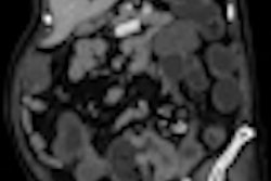
Both actuators were tested on the same volunteer on a 3-tesla clinical system (Verio, from Siemens Healthcare) using a single-shot echo planar imaging sequence with added motion encoding gradients, and data were analyzed using the complex inversion of the shear-wave algorithm. The group also tested the commercial Ehman actuator from the Mayo Clinic. Motion deflection tests were carried out on a gelatine phantom at various frequencies using a commercial accelerometer (Steval MKI005V1, STMicroelectronics).
The piezoelectric setup (75%) proved to be more powerful at higher frequencies. With the same setup at 40%, similar results were obtained compared with the commercial pneumatic actuator from Mayo Clinic, confirming the consistency of both actuators, the researchers stated.
"The custom-made pneumatic actuator was easy to install in the MR environment, and it was cheap compared to the other two setups. Despite proving to be less powerful and not so reliable as the other two setups, it still generated waves in phantom and liver experiments that were compared with the images obtained with the piezoelectric setup," they pointed out.
Wave images appeared more uniform using the piezoceramic device, suggesting that higher frequencies could still be employed with this system with reduced attenuation in the tissue of interest. However, for the pneumatic setup, in addition to lower amplitude irregular waves, the device should be restricted to lower vibration frequencies.
The team found that reconstructed shear stiffness maps appear smoother and more uniform using the piezoelectric system. Despite having noisier maps, the pneumatic system still produces results that are consistent with the results obtained with the piezoelectric setup.
"Although the pneumatic device offers an easier and faster system to integrate in the MR environment, the piezoelectric actuator can achieve higher frequencies and provide more reliable quantitative measurements. Nevertheless, other factors such as cost and complexity are a disadvantage with the piezoelectric system," they concluded.