Computer-aided detection (CAD) software can add additional benefits -- without too many false positives -- over double reading of low-dose chest CT examinations in a lung cancer screening environment, researchers from the Netherlands have found in research published in the October issue of European Radiology.
"Using a combination of CAD and nodule size cutoff in lung cancer screening improves the sensitivity of pulmonary nodule detection compared to that of double reading, without missing lung cancers," stated lead author Dr. Yingru Zhao of University Medical Center Groningen in Groningen, adding that CAD yielded 96.7% sensitivity for detecting potentially significant lung nodules and outperformed the 78.1% sensitivity produced by independent double readers.
Seeking to test the hypothesis that CAD can increase sensitivity for lung nodule detection beyond the results realized by double reading, the researchers sought to evaluate CAD's performance for finding nodules as a complementary tool in a large-scale, low-dose CT lung cancer screening study (Eur Radiol, October 2012, Vol. 22:10, pp. 2076-2084).
They randomly selected 400 cases out of the 4,280 baseline CTs from the Nederlands-Leuvens Longkanker Screenings Onderzoek (NELSON) trial. The CT scans were retrospectively evaluated by two independent readers with six years of experience and were then processed by CAD (LungCAD VB10A, Siemens Healthcare).
At least one finding was reported on 332 of the 400 baseline CT examinations. The readers and CAD system detected an aggregate of 1,667 findings (311 were found by double reading and CAD, 24 were found only by double reading, and 1,332 were only found by CAD).
Of these 1,667 findings, 1,516 (90.9%) could be excluded from further evaluation; 49.2% were excluded due to their small size (< 50 mm3), 37.6% were excluded as they were judged to be benign lesions, and 13.2% were excluded as they were determined to be nonlesions. The 151 findings that needed further evaluation were contributed by double reading and CAD in 113 (74.8%) of cases, by double reading only in five cases (3.3%), and only by CAD in 33 (21.9%) cases.
Performance in nodules > 50 mm3
|
"CAD was better in detecting most types of nodules, namely peripheral and nonperipheral nodules, solid nodules, intraparenchymal nodules, and spherical and nonspherical nodules," the authors wrote.
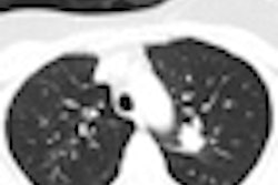
Readers struggled to detect vessel-attached nodules, finding only 11 (37.9%) of 29. Of the nodules found by CAD but undetected by the readers, 69.7% were attached nodules; 78.3% of the attached nodules were vessel-attached. Seven of 11 nonperipheral, vessel-attached nodules were missed by readers, but all 11 were detected by CAD.
Of the 33 nodules missed by the double readers but found by CAD at baseline, 24 were detected at three-month or one-year follow-up CT exams, according to the authors.
"Lung cancer was diagnosed in one solid intraparenchymal nodule, found to have grown at the second-year screening CT," the authors noted. "The baseline volume of this missed nodule was 160.7 mm3."
CAD had some misses of its own, failing to find one fissure-attached and two pleural-based nodules. Of the five nodules that were not detected by CAD, two were nonsolid or part-solid nodules. A solid pleural-based nodule with a volume of 217.8 mm3 was later diagnosed as cancer after it was found to be growing on the three-month follow-up CT exam.
As is often found with the technology, CAD also produced high false-positive rates. But using a nodule volume-cutoff threshold could sharply reduce that problem, the researchers found.
"Adding a nodule volume cutoff of 50 mm3 to CAD leads to nearly half the false-positive rate (1.9 versus 3.7 false-positive/CT) with an increase in positive predictive value," the authors noted.


![Examples of ultrasound findings and techniques. (A) Images in a 39-year-old male patient with a mass in the left thigh. The mass is heterogeneous on the B-mode US image (compared with the patient in D) and showed increased microvascularity (superb microvascular imaging [SMI]) and shear-wave elastography (SWE) values. Undifferentiated pleomorphic sarcoma was diagnosed at biopsy (with pleomorphic rhabdomyosarcoma in surgical specimen). (B) Images in an 18-year-old male patient with a mass in the left leg. The mass is hypoechoic on the B-mode image, with no other findings suggestive of malignancy. The lesion is in contact with the cortex of the tibia, which is slightly irregular. CT revealed a doubtful anteromedial tibial erosion. The microvascular study demonstrated high vascularization, suggestive of malignancy. Periosteal Ewing sarcoma was diagnosed with both histologic and immunohistochemical confirmation. (C) Images in a 69-year-old female patient with a lump growing on the outside of the left leg. Multiple SWE examinations were performed (please note the high values obtained in the measurements, whereas the color map highlights the stiffness relative to adjacent tissues). SMI showed areas of increased vascularization to target for sampling. Undifferentiated spindle cell sarcoma was diagnosed at biopsy, with residual leiomyosarcoma in the surgical specimen after neoadjuvant therapy. (D) Images in a 56-year-old female patient with a mass in the right thigh. The mass is heterogeneous at both B-mode ultrasound (similar to patient A) and MRI (coronal T2-weighted spectral attenuated inversion recovery [SPAIR]; T1-weighted pre-contrast and postcontrast imaging), which even shows uptake after the administration of paramagnetic contrast material, which is traditionally suggestive of malignancy. Low values at SMI and elastography are suggestive of benignity. Spindle cell lipoma was diagnosed at biopsy, with atypical spindle cell lipomatous tumor in the surgical specimen.](https://img.auntminnieeurope.com/mindful/smg/workspaces/default/uploads/2026/08/images-radiol250278fig2.APCFLSvX6p.jpg?auto=format%2Ccompress&fit=crop&h=100&q=70&w=100)


![Examples of ultrasound findings and techniques. (A) Images in a 39-year-old male patient with a mass in the left thigh. The mass is heterogeneous on the B-mode US image (compared with the patient in D) and showed increased microvascularity (superb microvascular imaging [SMI]) and shear-wave elastography (SWE) values. Undifferentiated pleomorphic sarcoma was diagnosed at biopsy (with pleomorphic rhabdomyosarcoma in surgical specimen). (B) Images in an 18-year-old male patient with a mass in the left leg. The mass is hypoechoic on the B-mode image, with no other findings suggestive of malignancy. The lesion is in contact with the cortex of the tibia, which is slightly irregular. CT revealed a doubtful anteromedial tibial erosion. The microvascular study demonstrated high vascularization, suggestive of malignancy. Periosteal Ewing sarcoma was diagnosed with both histologic and immunohistochemical confirmation. (C) Images in a 69-year-old female patient with a lump growing on the outside of the left leg. Multiple SWE examinations were performed (please note the high values obtained in the measurements, whereas the color map highlights the stiffness relative to adjacent tissues). SMI showed areas of increased vascularization to target for sampling. Undifferentiated spindle cell sarcoma was diagnosed at biopsy, with residual leiomyosarcoma in the surgical specimen after neoadjuvant therapy. (D) Images in a 56-year-old female patient with a mass in the right thigh. The mass is heterogeneous at both B-mode ultrasound (similar to patient A) and MRI (coronal T2-weighted spectral attenuated inversion recovery [SPAIR]; T1-weighted pre-contrast and postcontrast imaging), which even shows uptake after the administration of paramagnetic contrast material, which is traditionally suggestive of malignancy. Low values at SMI and elastography are suggestive of benignity. Spindle cell lipoma was diagnosed at biopsy, with atypical spindle cell lipomatous tumor in the surgical specimen.](https://img.auntminnieeurope.com/mindful/smg/workspaces/default/uploads/2026/08/images-radiol250278fig2.APCFLSvX6p.jpg?auto=format%2Ccompress&fit=crop&h=112&q=70&w=112)