As many as three-fourths of patients with chest pain could avoid invasive and costly procedures like invasive coronary angiography by undergoing noninvasive imaging tests first, according to investigators at the Evaluation of Integrated Cardiac Imaging (EVINCI) Consortium last week.
The prevalence of significant coronary artery disease in patients with chest pain is lower than expected in Europe, the investigators said at a meeting last week in Madrid.
Effective treatment of acute coronary syndromes and elimination of risk factors for coronary artery disease has reversed the prevalence of chronic disease and heart failure, the group said.
As a result, health systems are seeing more patients with suspected coronary artery disease who need effective diagnosis and treatment, but not necessarily invasive procedures. Only 40% of patients presenting with chest pain actually have obstructive disease, they noted.
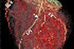
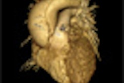
EVINCI's major goal was to assess the ability of noninvasive multimodality imaging (coronary CTA, SPECT, and PET) to diagnose the presence of coronary artery disease, determine if it primarily involves the major coronary arteries or the microvessels, and evaluate whether the disease is causing ischemia, which needs aggressive treatment, the consortium said in a statement.
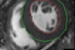
In addition, the effects of myocardial ischemia on ventricular function were assessed by either MRI or echocardiography during stress. Patients were followed up for a maximum of three years.
The EVINCI team enrolled 695 patients (ages 30-75 years, 38% women) with chronic chest pain and a 60% average probability of having coronary artery disease at 17 clinical centers in Europe.
After noninvasive screening, all patients underwent heart catheterization (coronary angiography and functional measurements) when appropriate as a reference method to define the presence, extent, and functional relevance of coronary disease, that is, its effect in limiting coronary flow reserve.
The different noninvasive strategies were compared for their diagnostic accuracy, and the costs and potential risks were monitored. This study design is in line with the push to perform comparative effectiveness research in Europe, the consortium said.
Knowing the extent and the functional relevance of the disease helps to determine whether treatment is likely to improve prognosis and quality of life, or whether it is unnecessary and can be avoided, the researchers said.
Preliminary results showed that in patients with stable angina or angina-like symptoms, the probability of significant coronary artery disease, based on clinical and stress ECG evaluation, is largely overestimated in current practice, the group said. As a result, many patients may receive unnecessary invasive procedures.
The average estimated probability of disease in the EVINCI population was 60%, while the actual prevalence of significant coronary disease, confirmed at heart catheterization and deserving revascularization, was only 25%, they said.
A final analysis of EVINCI data, expected in the next few weeks, will likely demonstrate that performing adequate noninvasive imaging screening of patients with suspected coronary artery disease could safely avoid invasive procedures in 75% the group stated.
Noninvasive cardiovascular imaging has a crucial role in identifying patients with significant obstructive coronary stenosis requiring revascularization. Noninvasive imaging will reduce the use of inappropriate invasive procedures and related costs, reducing risks for patients and identifying those who truly need revascularization, the consortium stated.


![Examples of ultrasound findings and techniques. (A) Images in a 39-year-old male patient with a mass in the left thigh. The mass is heterogeneous on the B-mode US image (compared with the patient in D) and showed increased microvascularity (superb microvascular imaging [SMI]) and shear-wave elastography (SWE) values. Undifferentiated pleomorphic sarcoma was diagnosed at biopsy (with pleomorphic rhabdomyosarcoma in surgical specimen). (B) Images in an 18-year-old male patient with a mass in the left leg. The mass is hypoechoic on the B-mode image, with no other findings suggestive of malignancy. The lesion is in contact with the cortex of the tibia, which is slightly irregular. CT revealed a doubtful anteromedial tibial erosion. The microvascular study demonstrated high vascularization, suggestive of malignancy. Periosteal Ewing sarcoma was diagnosed with both histologic and immunohistochemical confirmation. (C) Images in a 69-year-old female patient with a lump growing on the outside of the left leg. Multiple SWE examinations were performed (please note the high values obtained in the measurements, whereas the color map highlights the stiffness relative to adjacent tissues). SMI showed areas of increased vascularization to target for sampling. Undifferentiated spindle cell sarcoma was diagnosed at biopsy, with residual leiomyosarcoma in the surgical specimen after neoadjuvant therapy. (D) Images in a 56-year-old female patient with a mass in the right thigh. The mass is heterogeneous at both B-mode ultrasound (similar to patient A) and MRI (coronal T2-weighted spectral attenuated inversion recovery [SPAIR]; T1-weighted pre-contrast and postcontrast imaging), which even shows uptake after the administration of paramagnetic contrast material, which is traditionally suggestive of malignancy. Low values at SMI and elastography are suggestive of benignity. Spindle cell lipoma was diagnosed at biopsy, with atypical spindle cell lipomatous tumor in the surgical specimen.](https://img.auntminnieeurope.com/mindful/smg/workspaces/default/uploads/2026/08/images-radiol250278fig2.APCFLSvX6p.jpg?auto=format%2Ccompress&fit=crop&h=100&q=70&w=100)


![Examples of ultrasound findings and techniques. (A) Images in a 39-year-old male patient with a mass in the left thigh. The mass is heterogeneous on the B-mode US image (compared with the patient in D) and showed increased microvascularity (superb microvascular imaging [SMI]) and shear-wave elastography (SWE) values. Undifferentiated pleomorphic sarcoma was diagnosed at biopsy (with pleomorphic rhabdomyosarcoma in surgical specimen). (B) Images in an 18-year-old male patient with a mass in the left leg. The mass is hypoechoic on the B-mode image, with no other findings suggestive of malignancy. The lesion is in contact with the cortex of the tibia, which is slightly irregular. CT revealed a doubtful anteromedial tibial erosion. The microvascular study demonstrated high vascularization, suggestive of malignancy. Periosteal Ewing sarcoma was diagnosed with both histologic and immunohistochemical confirmation. (C) Images in a 69-year-old female patient with a lump growing on the outside of the left leg. Multiple SWE examinations were performed (please note the high values obtained in the measurements, whereas the color map highlights the stiffness relative to adjacent tissues). SMI showed areas of increased vascularization to target for sampling. Undifferentiated spindle cell sarcoma was diagnosed at biopsy, with residual leiomyosarcoma in the surgical specimen after neoadjuvant therapy. (D) Images in a 56-year-old female patient with a mass in the right thigh. The mass is heterogeneous at both B-mode ultrasound (similar to patient A) and MRI (coronal T2-weighted spectral attenuated inversion recovery [SPAIR]; T1-weighted pre-contrast and postcontrast imaging), which even shows uptake after the administration of paramagnetic contrast material, which is traditionally suggestive of malignancy. Low values at SMI and elastography are suggestive of benignity. Spindle cell lipoma was diagnosed at biopsy, with atypical spindle cell lipomatous tumor in the surgical specimen.](https://img.auntminnieeurope.com/mindful/smg/workspaces/default/uploads/2026/08/images-radiol250278fig2.APCFLSvX6p.jpg?auto=format%2Ccompress&fit=crop&h=112&q=70&w=112)