Using ECG-gated 320-detector-row CT, cardiac imaging researchers have developed a CT angiography (CTA) scan protocol that includes the heart and the entire aorta in four or five heartbeats -- all using prospective gating that reduces the radiation dose to reasonable levels considering the scan length, found a study published in European Radiology.
Using the wide-volume acquisition technique, the Beijing-based team performed 61 examinations successfully, delivering diagnostic image quality in the ascending aorta, aortic valve, and coronary arteries, and a mean radiation dose of 18 mSv to cover anatomic territory from the aortic arch to the aortic bifurcation, the group reported.
"Patients presenting with aortic diseases such as aortic aneurysm, penetrating aortic ulcer (PAU), aortic dissection (AD), and intramural hematoma (IMH) often share the same risk factors as coronary artery disease," noted Dr. Yu Li, Dr. Zhanming Fan, and colleagues from Capital Medical University (European Radiology, 4 June 2012, online first).
In patients with type A aortic dissection, rapid evaluation of the presence of coronary anomalies and atherosclerosis of the coronary arteries, and determining whether the dissection extends into the coronary vessels, is critical before surgery. For patients with symptoms suggesting aortic rupture and significant stenosis, "additional coronary CT angiography imaging would be required at the cost of increased potential risks from more contrast agents and radiation dose, increased cost, as well as delaying emergency aortic surgery," they wrote.
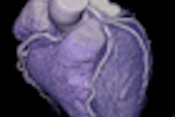
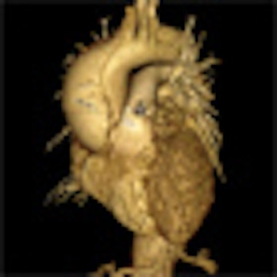
 Images of the whole aorta were acquired using a wide-volume protocol; each volume was scanned in a single heartbeat with 16-cm anatomic coverage. The second volume was positioned to cover the whole heart to ensure that all the segments of coronary arteries were in the same dataset. The table is stationary during each acquisition, and then moves to the next location for the next CT data acquisition. Patient is a 23-year-old woman with Marfan syndrome combined with coronary-pulmonary fistula, and a Stanford type A aortic dissection. All images courtesy of Dr. Yu Li.


For all the power of modern CT scanners to generate exquisitely detailed images of the vasculature, scanning the aorta without gating is risky because artifacts in the ascending aorta can simulate the appearance of a dissection flap, leading to an incorrect diagnosis of dissection. In their study, Li and colleagues aimed to investigate the feasibility of using a prospective ECG-gated wide-volume protocol in CTA of the whole aorta and coronary arteries (CA).
Between 2009 and 2010, 61 emergency department patients (47 men, 14 women; mean age 51.3 ± 18.9 years) underwent the whole-aorta scan following contrast administration but without beta-blockers. Exams were performed on a 320-detector-row CT scanner (Aquilion One, Toshiba Medical Systems) with patients in a supine position. Using a wide-volume protocol of four or five volumes, according to height, all exposures were acquired in a single volume.
For patients with heart rates below 70 bpm, data were acquired from 70% to 80% of the RR interval; 40% to 50% was used for patients with higher heart rates. Tube voltage and current were based on the patient's body mass index (BMI). The volumes were stitched together after acquisition to generate a CT dataset of the whole aorta. Images were initially reconstructed at fixed RR intervals, with additional reconstructions performed in 1% phases to achieve diagnostic images, Yi and colleagues explained.
Images were postprocessed on a workstation (Vitrea FX Workstation, Vital) by an experienced technologist who created 1-mm-thick axial and multiplanar reformations as well as volume-rendered and maximum-intensity projection images of the aorta. Multiplanar reconstructions of the angiographic view also were created for analysis of the coronary arteries. The images were reviewed by two experienced cardiac radiologists who were blinded to patients' clinical information.
Motion artifacts, including blurring and stair-step artifacts were assessed in the ascending aorta and aortic valve, and graded on a three-point scale, with 1 being good and 3 inadequate. Coronary arteries were evaluated on a four-point scale, with 4 being optimal.
All scans were successful with acceptable image quality in the ascending aorta, aortic valve (100%), and coronary arteries (94.4%), Yu and colleagues reported. The wide-volume technique was used to successfully image 78.7% (48/61) patients with five volumes and 21.3% (13/61) patients with four volumes, with a mean radiation dose of 18.42 ± 5.02 mSv. The mean heart rate was 85 ± 12.0 bpm, and the mean heart rate variability was 28.7 ± 14.5 bpm.
Stair-step artifacts between two adjacent datasets were seen in 10 patients, the authors said. Contrast enhancement greater than 300 HU was found in all measurements of the ascending aorta, descending aorta, and bifurcation of the aorta. The researchers achieved diagnostic image quality (scores 1 and 2) in all 61 patients in the evaluation of the whole aorta and the aortic root including sinus and valves, and there was excellent interobserver agreement on image quality grading (k = 0.815; 95% confidence interval: 0.60-1.00) for the ascending aorta and the aortic valves.
In all, 14 patients were diagnosed with aortic aneurysm and 35 with aortic dissection or intramural hematoma. Significant coronary artery stenosis was found in 12 patients.
"Type A aortic dissection with the involvement of the aortic root and extension around the aortic valve combined with aneurysmal dilatation of the aortic root was diagnosed in eight patients," the researchers wrote. In four of these cases, the right coronary artery also was involved.
A bicuspid valve was found in one patient, and normal valves were diagnosed in other patients. Twelve patients had greater than 50% stenosis in at least one vessel, including six of the 12 combined with aortic aneurysm, they wrote.
CTA of the aorta is a common imaging technique due to its speed and resolution, according to the researchers. But motion in the ascending aorta can be a problem without ECG gating. Using 320-detector-row CT, image quality is acceptable throughout, and for Stanford type A aortic dissection, CTA showed the relationship between the intimal flap, and the aortic valve and the coronary ostium "without obvious motion artifacts," they stated.
Fujioka et al found similar results in a study using 64-detector-row CT; however, heart rates were limited to 75 bpm or less, which is sometimes hard to achieve even with beta-blockers, they wrote. Some studies have shown that using ECG-gated sequential or high-pitch dual-source techniques produces good results in patients with high or unstable heart rates.
"The use of wide-volume CT techniques incorporating arrhythmia rejection software is unique to 320-detector-row CT systems," the researchers noted. "The arrhythmia rejection software recognizes an arrhythmia, stops the exposure, and waits for the next normal RR interval before acquiring the acquisition." In all, 6.8% of patients with high heart rates or arrhythmia had coronary segments with nondiagnostic image quality. Mean exposure time, 1.68 ± 0.14 seconds, is shorter than that achieved with 64-detector-row CT (29 ± 6 seconds), but similar to that achieved with high-pitch dual-source CT, they stated.
Among the limitations, the relatively high radiation dose of whole-aorta CTA is a concern; however the 320-detector-row technique produces longer anatomic coverage reported in previous triple-rule-out CTA studies with a similar radiation dose. And wide-volume acquisition with 320-detector-row CT eliminates the overbeaming effect.
The results were not correlated to angiography, the researchers noted. And using a heart-rate-based method to preset the exposure "may contribute negatively to coronary artery image quality because of heart rate accelerations (or decelerations) during data acquisition," they stated.
"We used an exposure window of 10% for single-heartbeat 320-row CTA, which will reduce the effective radiation dose when compared to full RR exposure and yield diagnostic images in greater than 95% of coronary arteries segments and all of the whole aorta and the aortic root (sinus and valves)," they wrote. "But the second generation of dual-source CT angiography of the thoracoabdominal aorta at a high pitch can deliver diagnostic depiction of the aortic root at a low radiation dose."
Prospective ECG-gated wide-volume protocol in CT angiography of the whole aorta may substantially lower the radiation dose while improving image quality of the ascending aorta and coronary arteries without beta-blockers in patients with suspected acute aortic diseases, the researchers concluded.
"For patients with aortic diseases, CTA of the whole aorta using a prospective ECG-gated wide-volume protocol has the potential to provide additional information about the coronary arteries and aortic valve with lower radiation exposure," Li wrote in an email to AuntMinnieEurope.com.
Next up, the project will continue with another scanner model. "Now we continue this study on the Definition Flash CT scanner [Siemens Healthcare] because the radiation dose is lower than the Aquilion One," Lee concluded.