Radiologists with limited experience in reading breast examinations may benefit significantly from using photon-counting tomosynthesis, leading researchers from Cambridge and Stockholm have found. In their study published in Radiology, two-view tomosynthesis outperformed 2D mammography for readers with the least amount of experience.
The value of annual mammography screening remains controversial, but for the most part radiologists agree on its utility. With the development of full-field digital mammography, even more cancers are detected, especially in women with dense breasts. The major drawback, however, is "the difficulty of finding and discerning a subtle change against the complex background of the glandular tissue, a particular issue for younger premenopausal women with a so-called dense glandular background pattern," wrote Dr. Matthew Wallis, a consultant radiologist and the director of the Cambridge and Huntingdon Breast Screening Service in the U.K. (Radiol, 24 January 2012, online first).
With tomosynthesis, low-dose images are obtained over a limited arc, and the projections are mathematically reconstructed into a series of sections with the hope that specificity and sensitivity will be improved. Initial work using tomosynthesis as an add-on to conventional 2D mammography suggested the potential for substantial improvements in specificity, the authors noted. They compared the diagnostic accuracy of 2D digital mammography with that of two-view (mediolateral and craniocaudal) and single-view (mediolateral oblique) tomosynthesis in an observer study involving two institutions: Cambridge and Capio St Göran's Hospital in Stockholm.
Ten accredited readers classified 130 women with breast density of 2 to 4 using 2D mammography and two-view tomosynthesis. Another 10 reviewed the same cases using 2D mammography but with single-view tomosynthesis.


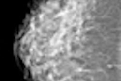
 A malignant spiculate mass in the right upper outer quadrant is seen better on photon-counting tomosynthesis. All images courtesy of Dr. Matthew Wallis.
For diagnostic accuracy, 2D mammography performed significantly worse than two-view tomosynthesis (average area under receiver operating characteristic curve [AUC] = 0.772 for 2D, AUC = 0.851 for tomosynthesis; p = 0.021). The researchers found significant differences for masses and microcalcification (p = 0.037 and 0.049, respectively). The difference in AUC between the two modalities was significant only for the five readers with the least amount of experience (p = 0.03 versus p = 0.25 for readers with 10 or more years of experience). No significant difference was seen in reader performance when 2D mammography was compared with single-view tomosynthesis.
The researchers also found that a two-view tomosynthesis examination takes almost twice as long to read as 2D mammograms.
"The role of tomosynthesis is still being clarified," Wallis wrote. "It has the potential to be an additional tool for the workup of screening-detected and clinical abnormalities."
The researchers acknowledge it would be tempting to suggest it's possible to perform screening by using single-view tomosynthesis, but at half the total radiation dose given that single-view tomosynthesis is performed at the same level as 2D mammography. "However, a number of smaller lesions were not visible on the single-view projection because they were not included on the image due either to their position in the breast or to poor technique, meaning that it would be important to test this proposition in a large real-life prospective study to ensure that equity was maintained," Wallis stated.


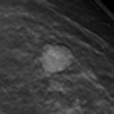
 Benign postoperative scarring. The distortion and trapping of the fat lobules and oil cysts are seen on both examinations but the photon-counting tomosynthesis offers better delineation and thus improves certainty of the less experienced reader.
Study limitations
Case selection could have been biased (the researchers collected a proportion of cases from screening assessment, which means they had already been detected at conventional mammography).
The researchers identified an outlier in the two-view tomosynthesis study arm whose AUC for the 2D mammographic images approached the guess line. However, they repeated the analysis with and without the reader, with similar results.
Tomosynthesis is a relatively new technique and all the readers are very experienced with 2D mammography. It is unrealistic to believe formal training in tomosynthesis can be a substitute for many years of experience.
This is an artificial study situation assessing single readers.
In Europe, large-volume readers offering double reading is predominant, so "great care needs to be taken in extrapolating the results to real life in two very different clinical environments," the researchers wrote. "When compared with single-view tomosynthesis, two-view tomosynthesis performs better than 2D mammography for less experienced readers regardless of radiologic feature, but care must be taken in overinterpreting results from this enriched test set study."
The study and detector development were funded by a European Union grant (LSHC-CT-2007-037642). Sectra provided the mammography equipment and employs two of the researchers. However, Wallis and two other researchers -- not employed by Sectra -- had full control of inclusion of all data, the information submitted for publication, and information that might present a conflict of interest.