If children diagnosed with cancer are old enough to understand the implications of radiation exposure, it may be wise to tell them to regard their imaging tests and treatments like a balancing act. Not enough radiation may fail to cure; too much could cause illness in the future.
Two radiologists from Italy have a similar perspective, and their judicious overview of the role of CT in pediatric oncology was published online 29 December in the European Journal of Radiology. The advantages of CT for treatment planning are the ability to image large anatomic areas rapidly with excellent spatial resolution and negligible motion artifacts. Also, unlike MRI exams, sedation of the patient is seldom required.
The ordering of CT scans should be kept to a minimum, based on specific diagnostic imaging guidelines. Multiphase studies should be avoided. MRI should be utilized if further characterization of a lesion with multiple phases is needed. When possible, ultrasound or MRI exams should be used for follow-up procedures to minimize additional high doses of radiation, according to Dr. Claudio Granata and Dr. Gianmichele Magnano of IRCCS Giannina Gaslini Hospital, University of Genoa.
CT protocols should be developed and consistently utilized. CT modalities need to be optimized to deliver the lowest possible amount of radiation dose exposure to obtain a diagnostic quality image. Image reconstruction should be used to reduce noise in images.
The authors discuss the use of CT for the following types of cancers:
Bone tumors: Bone lesions are common in children of all ages, but the vast majority are benign. For this reason, an x-ray should be the first diagnostic imaging exam performed. If a cancerous tumor is suspected, CT images can help detect subtle bone changes and precisely locate a lesion in the periosteal, cortical, or medullary bone. The authors point out that CT has a limited role in staging the treatment for bone tumors and that MRI is the recommended modality.
Hepatoblastoma: This liver tumor accounts for approximately 2% of all pediatric cancers, usually occurring when patients are younger than age 5. Either CT or MRI should be used for preoperative staging. If a CT exam is performed, it should be limited to a single portal venous phase, as multiple pre- and postcontrast phases usually do not add useful information for diagnosis and staging, the authors reported. The most important information from a CT exam is tumor extension within the liver, involvement of the portal vein, inferior vena cava, and/or hepatic veins, they wrote. The presence of extrahepatic diseases and distant metastases also should be determined. A Doppler ultrasound study should be performed to confirm vessel involvement if identified on CT images. A follow-up CT or MRI exam is needed following a patient's completion of preoperative chemotherapy.
Lymphoma: Approximately 15% of all childhood cancers are lymphomas. A definitive diagnosis should be made through histologic analysis, but a contrast-enhanced CT of the chest, abdomen, and neck should be performed. This exam will provide a detailed evaluation of the mediastinum, chest wall, pulmonary parenchyma, pleura, and pericardium; involvement of the cervical nodes of the Waldeyer ring; as well as enlarged abdominal and pelvic nodes. Involvement of the liver, spleen, kidneys, mesentery, and peritoneum also will be easier to determine.
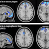
With respect to staging, the use of PET or PET/CT has modified treatment plans for up to 25% of adult cases, but little has been published about this subject with respect to pediatric cases. Similarly, there is little published information about its ability to define the involved regions requiring radiotherapy in children, although it seems to be more accurate than CT or MR images for adults. Because of its high effective radiation dose of 4.3 to 6.2 mSv in children, the use of PET/CT is controversial.
Neuroblastoma, ganglioneuroblastoma, and ganglioneuroma: These are the most common extracranial solid tumors in pediatric patients, and half of these tumors have reached a metastatic stage by the time of diagnosis. To determine if locoregional tumors are resectable or unresectable, and to identify the stage of the cancer, imaging is essential.
The authors advise that the primary tumor should be measured with 3D orthogonal measurements. The presence or absence of imagine-defined risk factors identified by the International Neuroblastoma Risk Group Staging System (INRGSS) should also be verified according to nine tumor sites and reported. The authors provided a list of specific terms that should be used to describe the relationship between the tumor mass and vital structures that if resected would impair normal function.
When bone or bone marrow is suspected of having metastatic disease, iodine-123 metaiodobenzylguanidine scintigraphy should be performed before tumor resection rather than a CT scan. It is considered the gold standard for bone and bone marrow assessment.
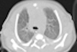
Pulmonary metastases: CT images are the reference standard for detecting pulmonary nodules or other abnormal findings. But they may not indicate if nodules are malignant or benign, and this issue is controversial. A chest x-ray also is recommended, depending upon other types of malignancies a pediatric patient may have.
Rhabdomyosarcoma: This is the most common pediatric soft-tissue sarcoma, usually located in the head, neck, genitourinary system, and limbs. An MRI exam is recommended by the European Pediatric Soft Tissue Sarcoma Study Group, but a CT scan of the abdomen and pelvis is recommended if the disease is found in the abdomen, lower limbs, or testes to assess lymph nodes and determine if it has spread to the liver.
Wilms' tumor: This malignant renal tumor is the most common one, but about 90% of children who have it survive. If ultrasound leads to diagnosis of a suspected Wilms' tumor, this should be verified with a CT or MRI exam. Because these patients tend to be younger than age 5, CT is the preferred modality.