Danish researchers have found that FDG-PET/CT can provide beneficial information on the possible malignancy of ovarian tumors and help gynecologists determine the correct course of treatment for patients.
They presented their study at the recent Society of Nuclear Medicine (SNM) annual meeting in Montreal, and also noted FDG-PET/CT can detect additional tumors, which leads to patient referrals to the appropriate medical specialist.
Lead study author Majbritt Frost, a research technologist at Aalborg Hospital in Aalborg, Denmark, noted that the ability to detect ovarian cancer expands hybrid molecular imaging in the field of gynecology.
Molecular imaging already is beneficial for the staging of cervical cancer, and continuing research may one day open the door for the staging of endometrial and other cancers for women, she added.
Adnexal masses
The purpose of the Danish study was to evaluate the accuracy of preoperative FDG-PET/CT in predicting malignancy in patients with adnexal masses compared with clinical pathological staging. Adnexal masses are lumps in the tissue of the adnexa or connected structures of the uterus. The masses most commonly affect the ovaries and fallopian tubes, but they can also develop within supporting tissues.
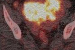
Clinicians imaging patients suspected of malignant tumors can see where cancerous cells are hypermetabolizing FDG and accurately predict whether a mass is malignant, cancerous but stable, or benign. FDG-PET imaging ascertains whether lumps are malignant because cancerous tissues show more metabolic activity than normal cells.
The study included a total of 104 patients with a mean age of 62 years, ranging from age 32 to 89. Presenting masses with the potential for ovarian cancer were imaged using PET/CT with FDG prior to surgery. Researchers then compared the results of preoperative FDG-PET/CT findings with the intraoperative and histopathological findings consecutively from January 2008 to April 2010.
Results of the scans were classified as either benign or malignant and were then compared with surgical findings.
Tumor detection
Preliminary results found that FDG-PET/CT was able to successfully detect 84% of benign and malignant tumors for these patients. In addition, FDG-PET/CT's description of malignant and benign was correct in 86% of patients.
FDG-PET/CT demonstrated areas of abnormal metabolic activity highly suspicious for malignant tumors in 60 patients (58%). Tumors were considered benign on FDG-PET/CT in 44 (42%) of 104 patients. Histopathology showed malignant tumors in 53 patients (51%) and benign tumors in 51 patients (49%). Thus, FDG-PET/CT scans showed a sensitivity of 91% and specificity of 76%.
Based on the results, Frost and colleagues concluded that FDG-PET/CT "can be useful for preoperative differentiation between benign and malignant adnexal masses."
In addition, as a whole-body examination with intravenous contrast, FDG-PET/CT gives useful information of metastasis, extent of malignant diseases, and other concomitant pathologies.


![Overview of the study design. (A) The fully automated deep learning framework was developed to estimate body composition (BC) (defined as subcutaneous adipose tissue [SAT] in liters; visceral adipose tissue [VAT] in liters; skeletal muscle [SM] in liters; SM fat fraction [SMFF] as a percentage; and intramuscular adipose tissue [IMAT] in deciliters) from MRI. The fully automated framework comprised one model (model 1) to quantify different BC measures (SAT, VAT, SM, SMFF, and IMAT) as three-dimensional (3D) measures from whole-body MRI scans. The second model (model 2) was trained to identify standardized anatomic landmarks along the craniocaudal body axis (z coordinate field), which allowed for subdividing the whole-body measures into different subregions typically examined on clinical routine MRI scans (chest, abdomen, and pelvis). (B) BC was quantified from whole-body MRI in over 66,000 individuals from two large population-based cohort studies, the UK Biobank (UKB) (36,317 individuals) and the German National Cohort (NAKO) (30,291 individuals). Bar graphs show age distribution by sex and cohort. BMI = body mass index. (C) After the performance assessment of the fully automated framework, the change in BC measures, distributions, and profiles across age decades were investigated. Age-, sex-, and height-adjusted body composition reference curves were calculated and made publicly available in a web-based z-score calculator (https://circ-ml.github.io).](https://img.auntminnieeurope.com/mindful/smg/workspaces/default/uploads/2026/05/body-comp.XgAjTfPj1W.jpg?auto=format%2Ccompress&dpr=2&fit=crop&h=167&q=70&w=250)


