Powered by the advances in acquisition technology and image postprocessing, CT has emerged as a critical validation tool for oncologists in the assessment of tumors. Yet the radiology department has an image problem with oncologists, according to Dr. Philippe Grenier, who leads the department of diagnostic radiology at the Hôpital Pitié-Salpêtrière in Paris.
"The reports we send assessing the response of tumors to chemotherapy are inconsistent and often incomprehensible, and when an oncologist calls us, he learns no two radiologists have the same reading of the same images," he said. "Oncologists do not have a culture that includes medical imaging, and we have not done a very good job of building our credibility with them."
For the past eight months, Grenier's team has studied the potential for computer-assisted detection (CAD) software in second readings of CT exams, specifically testing its capabilities for exams of lung cancer patients following chemotherapy.
"It is nothing less than fantastic," said Grenier, past president of the Fleischner Society and president of the European Society of Thoracic Imaging.
He is convinced that CAD software is accelerating both the quality and the speed of follow-up CT readings in oncology.
"100% of tumors are imaged today, and 80% of this work is done with CT," Grenier said. "Now to compare tumor progression, we can click and voilà , the software shows the targeted nodules with variations in volumes from a previous scan."
CT is critical to the initial detection of a tumor and the supporting diagnosis, while it is also key to guiding biopsies and tumor treatment procedures. CT has also proved effective in follow-up exams that within the first three months of chemotherapy can indicate whether the treatment is working.
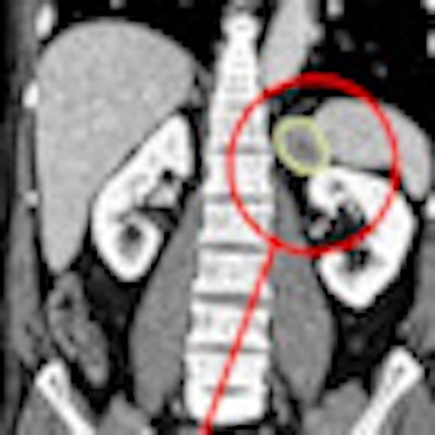
Computer-assisted follow-up exam of chemotherapy patient comparing prior and current coronal views. Changes to the target tumor are displayed and measured in an axial zoom view. Image courtesy of Median Technologies.Radiologists should be very pleased with CAD as it increasingly brings standardization to reporting, optimization of measurements, and consensus on methods for comparing different examinations, he said. The keen interest of pharmaceutical companies in quantifiable results for effectiveness of chemotherapies will provide a wealth of clinical evidence for radiologists to review and debate as they consider emerging standards.
Standardization should help convince oncologists about the value of imaging for assessing treatment, and will ensure radiologists improve the quality of their reporting.
"There is a serious problem with reproducibility in exams for tumor response with radiologists taking different measures for diameter and volumes," said Dr. Olivier Lucidarme, radiologist at Pitié-Salpêtrière who is leading the investigations into CAD software from Median Technologies of Valbonne, France.
International standards set a 20% increase in volume as the threshold for saying the tumor is growing, he explained. With two different sets of measures from the same tumor, the risk of error increases in reporting, whether a patient is responding or not to a treatment. The automation of second readings is also assisting in oncology therapy follow-up.


![Examples of ultrasound findings and techniques. (A) Images in a 39-year-old male patient with a mass in the left thigh. The mass is heterogeneous on the B-mode US image (compared with the patient in D) and showed increased microvascularity (superb microvascular imaging [SMI]) and shear-wave elastography (SWE) values. Undifferentiated pleomorphic sarcoma was diagnosed at biopsy (with pleomorphic rhabdomyosarcoma in surgical specimen). (B) Images in an 18-year-old male patient with a mass in the left leg. The mass is hypoechoic on the B-mode image, with no other findings suggestive of malignancy. The lesion is in contact with the cortex of the tibia, which is slightly irregular. CT revealed a doubtful anteromedial tibial erosion. The microvascular study demonstrated high vascularization, suggestive of malignancy. Periosteal Ewing sarcoma was diagnosed with both histologic and immunohistochemical confirmation. (C) Images in a 69-year-old female patient with a lump growing on the outside of the left leg. Multiple SWE examinations were performed (please note the high values obtained in the measurements, whereas the color map highlights the stiffness relative to adjacent tissues). SMI showed areas of increased vascularization to target for sampling. Undifferentiated spindle cell sarcoma was diagnosed at biopsy, with residual leiomyosarcoma in the surgical specimen after neoadjuvant therapy. (D) Images in a 56-year-old female patient with a mass in the right thigh. The mass is heterogeneous at both B-mode ultrasound (similar to patient A) and MRI (coronal T2-weighted spectral attenuated inversion recovery [SPAIR]; T1-weighted pre-contrast and postcontrast imaging), which even shows uptake after the administration of paramagnetic contrast material, which is traditionally suggestive of malignancy. Low values at SMI and elastography are suggestive of benignity. Spindle cell lipoma was diagnosed at biopsy, with atypical spindle cell lipomatous tumor in the surgical specimen.](https://img.auntminnieeurope.com/mindful/smg/workspaces/default/uploads/2026/08/images-radiol250278fig2.APCFLSvX6p.jpg?auto=format%2Ccompress&fit=crop&h=100&q=70&w=100)


![Examples of ultrasound findings and techniques. (A) Images in a 39-year-old male patient with a mass in the left thigh. The mass is heterogeneous on the B-mode US image (compared with the patient in D) and showed increased microvascularity (superb microvascular imaging [SMI]) and shear-wave elastography (SWE) values. Undifferentiated pleomorphic sarcoma was diagnosed at biopsy (with pleomorphic rhabdomyosarcoma in surgical specimen). (B) Images in an 18-year-old male patient with a mass in the left leg. The mass is hypoechoic on the B-mode image, with no other findings suggestive of malignancy. The lesion is in contact with the cortex of the tibia, which is slightly irregular. CT revealed a doubtful anteromedial tibial erosion. The microvascular study demonstrated high vascularization, suggestive of malignancy. Periosteal Ewing sarcoma was diagnosed with both histologic and immunohistochemical confirmation. (C) Images in a 69-year-old female patient with a lump growing on the outside of the left leg. Multiple SWE examinations were performed (please note the high values obtained in the measurements, whereas the color map highlights the stiffness relative to adjacent tissues). SMI showed areas of increased vascularization to target for sampling. Undifferentiated spindle cell sarcoma was diagnosed at biopsy, with residual leiomyosarcoma in the surgical specimen after neoadjuvant therapy. (D) Images in a 56-year-old female patient with a mass in the right thigh. The mass is heterogeneous at both B-mode ultrasound (similar to patient A) and MRI (coronal T2-weighted spectral attenuated inversion recovery [SPAIR]; T1-weighted pre-contrast and postcontrast imaging), which even shows uptake after the administration of paramagnetic contrast material, which is traditionally suggestive of malignancy. Low values at SMI and elastography are suggestive of benignity. Spindle cell lipoma was diagnosed at biopsy, with atypical spindle cell lipomatous tumor in the surgical specimen.](https://img.auntminnieeurope.com/mindful/smg/workspaces/default/uploads/2026/08/images-radiol250278fig2.APCFLSvX6p.jpg?auto=format%2Ccompress&fit=crop&h=112&q=70&w=112)