Researchers from the Netherlands used an automated coronary artery motion model to analyze cardiac function from electrocardiogram-gated coronary CT angiography (CTA) data. The method promises to improve cardiac function analysis and reconstruction interval selection, and even provide new information on coronary artery motion at different heart rates.
The prospective study, presented at the 2009 RSNA meeting in Chicago, examined 31 patients with known or suspected coronary artery disease who were referred for coronary CTA using a dual-source CT system (Somatom Definition, Siemens Healthcare, Erlangen, Germany).
The purpose of the study was to extract coronary artery dynamics over the entire cardiac cycle from derivation of electrocardiogram (ECG)-pulsed and gated CTA data, and analyze cardiac function from coronary CTA data, said Coert Metz, a Ph.D. student specializing in biomedical image analysis at Erasmus Medical Center in Rotterdam, Netherlands.
The heart was scanned in a single breath-hold using a heart-rate-dependent ECG-pulsing protocol, and without the use of beta-blockers, Metz said. Data were reconstructed using a slice thickness and reconstruction increment of 3.0/1.5 mm, at 5% steps from 0% to 95% of the RR interval.
"One of the main coronary arteries was annotated from one of three groups, either the [left anterior descending (LAD)], [left circumflex (LCX)], or [right coronary artery (RCA)]," Metz said. "They were annotated at both 30% and 60% of the RR interval, and then we derived the motion of the coronary arteries using an automated nonrigid co-registration method."
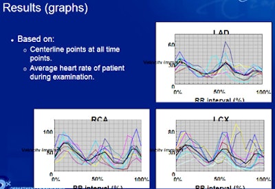
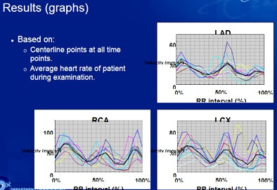
Beginning with, for example, a 60% RR image, the registration method was used to align the other image types to it, Metz said. Then the coronary centerline in the reference image was annotated and the centerline warped to the other time points. Displacement and average velocities were calculated from the centerlines, as well as the co-registration result and the average heart rate of the patients.
"In this way, we derived the 4D model of the coronary arteries for the patient, and we can do data measurements," he said. Intervals at systole and diastole with the least coronary motion were calculated automatically.
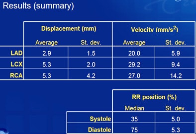
For LAD, results showed displacement of around 3 mm and average velocity of 20 mm/sec2 over the length of the artery, he said. For LCX, displacement was 5.3 mm with velocity of 29 mm/sec2, and for RCA displacement was also 5.3 mm with average velocity of 27 mm/sec2.
"We are quite happy with the results as they are similar to earlier studies," he said. The results showed that "velocity and displacement of the coronary arteries can be derived from ECG-gated, pulsed CTA data using image registration algorithms."
 |
| Results show average displacement and velocity of coronary arteries from CTA data for LAD, LCX, and RCA. RCA data show a higher standard deviation. All data courtesy of Coert Metz. |
 |
The standard deviations for displacement of the LAD and LCX arteries were low, suggesting adequate analysis, but higher standard deviations for the RCA (4.2) suggest that the model "may not be good enough" yet, Metz said. Analysis of the RCA is generally problematic in coronary CTA studies.
Future work will aim to determine whether semiautomatic centerline extraction can reduce user interaction time, and the group will also explore advanced analysis of cardiac function and motion, Metz said. "For example, it would be really interesting to investigate the relationship between the heart rate of the patient and the velocity of the coronary arteries," he said.
Another interesting topic to investigate is "how we can use this information to develop imaging protocols that can actually reduce motion artifacts," Metz told AuntMinnie.com.
In questions after the talk, some attendees were enthusiastic about the potential of the technique to better evaluate difficult artery segments, while others considered the velocity measurements inadequate.
"It's really not appropriate to use standard deviations in your analysis of the velocities, because the distribution of velocities over time is not bell-shaped, but severely asymmetric," commented one audience member.
In an e-mail to AuntMinnie.com, Metz said he agreed with the criticism.
"For future research, it would ... make more sense to investigate the velocity locally -- for example, per vessel segment or even compared to the distance from the ostium," Metz wrote. "What numbers to report depends also on the application. In this work, we just reported averages and standard deviation because we wanted to summarize the results in some way."
By Eric Barnes
AuntMinnie.com staff writer
January 11, 2010
Related Reading
CAD takes on arteries with coronary stenosis detection, November 19, 2009
Automated segmentation could improve fetal heart echo, November 6, 2009
CARS report: Fuzzy theory yields sharper CTA images, June 27, 2008
3D development outpaces facilities' readiness, June 23, 2008
Video game technology speeds beating-heart surgery, June 9, 2008
Copyright © 2010 AuntMinnie.com


