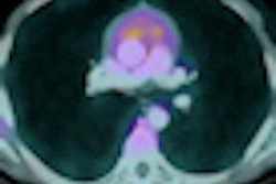
NEW YORK (Reuters Health), Aug 17 - Use of F-18 FDG-PET imaging can reduce the rate of futile laparotomies in patients with colorectal liver metastases from 45% to 28%, according to a report in the July issue of the Journal of Nuclear Medicine.
"This is the first and only randomized controlled clinical trial on the efficacy of FDG-PET to select patients with liver metastases from colorectal cancer for liver surgery," senior author Dr. Wim J. G. Oyen, from Radboud University Nijmegen Medical Centre, the Netherlands, told Reuters Health.
"The biggest finding," Dr. Oyen added, "is that FDG-PET detects additional disease that deems liver surgery futile in one in six patients, who would have been taken to surgery when CT alone was used. Furthermore, the addition of FDG-PET in every patient did not result in an increase of overall health care costs."
The results stem from a study of 150 patients with colorectal liver metastases who were randomly assigned to preoperative evaluation with CT alone or combined with FDG-PET. The subjects were followed for at least three years after randomization.
Futile laparotomy, the main outcome measure, was defined as a laparotomy that did not lead to complete tumor removal, showed benign disease, or that did not improve disease-free survival by longer than six months.
Thirty-four patients (45%) in the control group had a futile laparotomy compared with 21 (28%) in the PET group, a risk reduction of 38% (p = 0.042).
"Every patient with liver metastases from colorectal cancer to whom liver surgery is offered, should ask for FDG-PET before the final decision to actually go ahead with liver surgery is made," Dr. Oyen emphasized.
"Future research will focus on the role of neoadjuvant chemotherapy and the role of novel surgical procedures such as radiofrequency ablation of tumors," he added.
By Anthony J. Brown, M.D.
J Nucl Med 2009;50:1036-1041.
Last Updated: 2009-08-17 10:50:04 -0400 (Reuters Health)
Related Reading
FDG-PET helps assess recurrent colon cancer, January 19, 2009
FDG-PET detects recurrence of colorectal cancer early, April 3, 2008
Copyright © 2009 Reuters Limited. All rights reserved. Republication or redistribution of Reuters content, including by framing or similar means, is expressly prohibited without the prior written consent of Reuters. Reuters shall not be liable for any errors or delays in the content, or for any actions taken in reliance thereon. Reuters and the Reuters sphere logo are registered trademarks and trademarks of the Reuters group of companies around the world.