CHICAGO (Reuters), Jul 8 - An advanced type of MRI scan may help doctors more accurately diagnose a severe form of endometriosis, researchers said on Tuesday, potentially allowing some women to avoid invasive pelvic surgery.
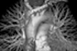
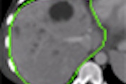
They said a new kind of magnetic resonance imaging scan called 3 tesla, or 3T MRI, helped doctors rule out deep endometriosis in 93% of young women studied.
Endometriosis is a chronic disease that results when uterine tissue, called endometrium, grows outside the uterus, causing pain in the pelvis and lower back, painful menstrual cramps, fatigue, and infertility. It affects 5 million American women.
In most cases, doctors can remove excess tissue using laparoscopic surgery, in which a lighted instrument is inserted through a small incision.
But in women with deep endometriosis, in which uterine tissue attaches itself to other organs such as the ovaries, fallopian tubes, and colon, excess tissue must be removed through a large incision in the abdomen, a riskier procedure that requires longer follow-up.
A team lead by Dr. Nathalie Hottat of the Universite Libre de Bruxelles in Brussels, Belgium, wanted to see if the high-tech MRI scanners could help surgeons better predict which women needed more invasive surgery.
Hottat's team performed MRI scans on 41 women ages 20 to 46 with suspected endometriosis prior to surgery.
They found MRI accurately diagnosed 26 of 27 cases of deep endometriosis, helping doctors to plan their surgery, the team reported in the journal Radiology.
The scans were highly accurate at determining which women would benefit from less-invasive surgery, ruling out deep endometriosis in 93% of cases and allowing surgeons to perform a less invasive laparoscopic procedure.
Source: Radiology, online July 7, 2009.
Last Updated: 2009-07-07 16:29:06 -0400 (Reuters Health)
Related Reading
3-tesla MRI sheds light on shading sign in endometriosis, June 8, 2007
Copyright © 2009 Reuters Limited. All rights reserved. Republication or redistribution of Reuters content, including by framing or similar means, is expressly prohibited without the prior written consent of Reuters. Reuters shall not be liable for any errors or delays in the content, or for any actions taken in reliance thereon. Reuters and the Reuters sphere logo are registered trademarks and trademarks of the Reuters group of companies around the world.