VIENNA - Even with electrocardiographic gating, CT angiography cannot confidently visualize subtle aortic tears of the ascending aorta, say researchers from Johannes Gutenberg University. Transesophageal echocardiography (TEE) may be a better option in some patients.
Diagnosing smaller intimal tears without hematoma or an intimal flap remains a challenge in patients presenting with symptoms of acute aortic syndrome but without significant internal hematoma, said Dr. Sebastian Schotten in a presentation on Thursday at the European Congress of Radiology (ECR).
"These lesions may progress, at least in some cases, to classic dissection or aortic rupture," Schotten said.
Among the few references to intimal tears of the ascending aorta in the literature, "MRI or nongated CT fails to make the diagnosis; however, gated CT has not yet been evaluated. Therefore, the purpose of our study was to assess the value of gated CT for subtle dissections of the ascending aorta, and correlate these results with intraoperative findings and TEE," he said.
The retrospective study included 32 patients with suspected Stanford type A dissection. All patients preoperatively underwent contrast-enhanced electrocardiogram (ECG)-gated CT angiography on a 64-detector-row scanner (Brilliance 64, Philips Healthcare) (n = 30) or a 256-detector-row scanner (iCT 256, Philips Healthcare) (n = 2).
Two readers working in consensus retrospectively analyzed the images, focusing on detecting intramural hematomas, pericardial and pleural effusions, mediastinal hemorrhages, and intimal flaps or tears. The investigators correlated the imaging findings with intraoperative findings and also with histopathology.
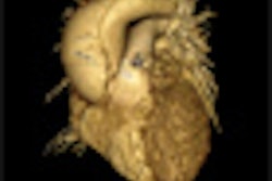
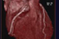
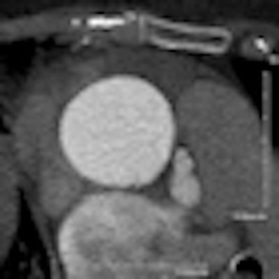
"ECG gating allows motion-free images of the aortic lumen ... in contrast to nongated CT," where image quality is degraded by motion artifacts, Schotten said.
Histopathology showed dissection of the ascending aorta with an intimomedial flap in 26 (81%) of 32 patients, an intramural hematoma in two (6%) patients, and a limited intimal tear in the ascending aorta in four (13%) patients.
"Intraoperatively, four patients were found to have limited intimal tears in the ascending aorta, and of these, in only two patients was the dissection revealed by CT," Schotten said. "These may range from very limited tears in the aortic wall to deeper and more extended tears with subsequent bleeding in the adventitia, but without classic dissection."
 |
| At ECG-gated cornary CTA, a small defect can be seen in the aortic wall of a 74-year-old woman referred for suspected pulmonary embolism. The lesion was found to correspond to a 12-mm supercoronary tear in the aortic wall at surgery. Image courtesy of Dr. Sebastian Schotten. |
ECG gating does limit motion artifacts of the aortic root and ascending aorta, and as a result it improves the identification of small intimal injuries, Schotten said. However, even with ECG gating not all subtle aortic dissections could be identified in the study.
"We can definitely conclude that subtle dissections are a challenge with even state-of-the-art diagnostic imaging," he said. TEE has also been shown to detect those lesions and therefore should be performed in patients with persistent symptoms and a negative CT scan.
Because TEE was performed in many of the patients, the group next plans to correlate those findings with CT to compare the accuracy of the two modalities, he said.
In response to a question from the audience, Schotten said there was no difference between the quality of the 64- and 256-detector-row images. Another audience member asked if every patient with such a subtle tear should be referred for surgery, because in some cases patients have been followed and the tears have resolved.
Schotten said the data are insufficient on how many small tears of the ascending aorta progress to classic dissection. "But in a center like ours where you have many patients operated on for the ascending aorta, and you have a low frequency of surgical complications and mortality, I think, yes, you should operate," he said.