In the field of ear, nose, and throat (ENT) medicine, the applications of artificial intelligence (AI) are numerous. They are growing particularly fast in the world of otology.
A recent inventory from the American ENT Academy found 10% of applications related to AI in this area of imaging, 13% related to hearing aids, 11% with a speech recognition aid, 29% related to the diagnosis and management of vestibular disorders, 24% related to prediction of the evolution of neurosensory deafness, and 13% to the analysis of auditory brainstem responses.
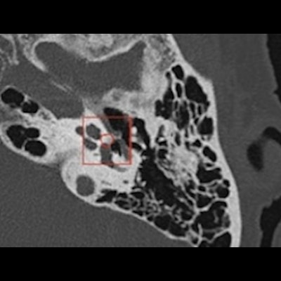
 45-year-old woman with predominantly right-sided unilateral conductive hearing loss. Double oblique view of the stirrup. Stapedial otosclerosis with isolated isodense filling of the fissula ante fenestram detected by the Oto-Med recognition tool. Courtesy of
 e-Quotidien, daily newspaper of Journées Francophones de Radiologie (JFR).
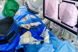
Regarding imaging, deep learning already allows clinical applications ranging from the automatic segmentation of anatomical structures, recognition of small pathologies such as otosclerosis, through to medical education, and even surgical planning with, in particular, facial nerve tracking. For otosclerosis, real performance results are at least equivalent to expert radiologists, even at stages I and II, also allowing differential diagnoses such as stapedial malformation or chronic otitis.
In a study using the algorithm developed using Cleverdoc tools and led by Oto-Med, the results of external validations of 124 cases studied on multibrand CTs, with otosclerosis confirmed surgically (Figure 2), were 96.6%, 95.3%, 95%, and 97% respectively for sensitivity, specificity, positive predictive value (PPV), and negative predictive value (NPV). The other tools developed relate to malformative and cholesteatoma pathologies and vestibular MRI. The future also lies in the integration of other biomarkers, whether clinical, biological, electrophysiological, anatomopathological, or genomic, to improve patient care.
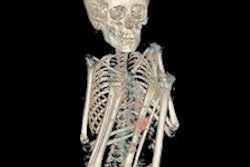
 Oto-Med / Surgery agreement in a patient operated on for left stapedial otosclerosis. Figure courtesy of Dr. Arnaud Attyé, Grenoble University Hospital Center, France.
It is also the integration of these technologies into everyday tools and in particular into PACS that will allow their development and easy routine use. This is crucial for the acceptance of these new tools. It is necessary to be able to access the analysis without changing environment and with a maximum response time of no more than a minute for routine clinical use.
We also insist, as with all other organ specialties, on the need for backup and archiving with data classification, in particular CT and MRI. The stakes have never been so critical for maintaining our independence and for developing and using these algorithms without forgetting the prime notions of ethics and regulations. As with any medical procedure, information and consent by the patient are necessary. They are the essential foundation for any algorithm development. The future is exciting and there are already many tools available. To prepare ourselves, our vigilance and our critical thinking should help us choose the right partners.
The AI revolution is underway. Its development, currently favored in settings with high medical resources, is primarily intended to facilitate quality care with more limited human resources. The applications are multiple, diagnostic, and therapeutic. Let us unite in order to develop these tools collaboratively, and ideally, with our organ societies, in this case CIREOL, (Collège d'Imagerie pour la Recherche et l'Enseignement en ORL), imaging college for ENT research and teaching.
Dr. Loïc Gaillandre, is a radiologist at C.L.I.M.A.L. medical imaging center, Lille, France.
Editor's note: This is an edited version of a translation of an article published in French online by the daily newspaper, e-Quotidien, of Journées Francophones de Radiologie (JFR). Translation by Frances Rylands-Monk. To read the original version, go to the JFR website.