PET/CT has no rivals when it comes to the detection and surveillance of cardiac metastases in well-differentiated gastroenteropancreatic neuroendocrine tumors (GEP-NETs) as well as guiding clinical management, according to research presented at the annual congress of the European Association of Nuclear Medicine (EANM 2020).
"Chest CT demonstrates poor sensitivity for detection of cardiac metastasis, with cardiac MRI performing much better. Still, neither CT nor MRI is as sensitive as [gallium-68 (Ga-68)] DOTATATE PET/CT for detection of neuroendocrine cardiac metastasis," noted Dr. John Renfrew and colleagues from the Mayo Clinic in Phoenix, Arizona, U.S.
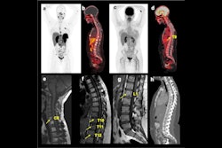
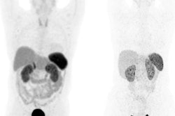
 Maximum intensity projection image of Ga-68 DOTATATE PET demonstrates focally increased tracer uptake in a metastasis involving the interventricular septum (arrow). Low-grade uptake seen within the bilateral inguinal lymph nodes was reactive. All images courtesy of Dr. John Renfrew, Mayo Clinic in Arizona.
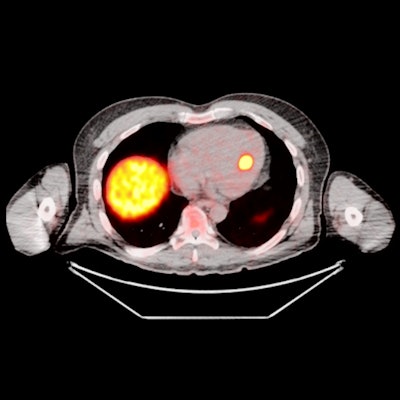
![Axial Ga-68 DOTATATE PET/CT image for the same patient demonstrates the focal tracer uptake localized to the interventricular septum (maximum standardized uptake value [SUVmax] = 24).](https://img.auntminnieeurope.com/files/base/smg/all/image/2020/10/ame.2020_10_27_16_51_9149_2020_10_27_mol_insider_Renfrew_figure2.png?auto=format%2Ccompress&dpr=2&fit=max&q=70&w=700) Axial Ga-68 DOTATATE PET/CT image for the same patient demonstrates the focal tracer uptake localized to the interventricular septum (maximum standardized uptake value [SUVmax] = 24).
Axial Ga-68 DOTATATE PET/CT image for the same patient demonstrates the focal tracer uptake localized to the interventricular septum (maximum standardized uptake value [SUVmax] = 24).Evidence from the Mayo Clinic confirms that the prevalence of cardiac metastasis is associated with high tumor burden, and an average of 2.7 other organ systems are also involved when these metastases are seen. Further, patients typically demonstrate a Krenning score of 3-4 when they have cardiac metastases.
Rare entities
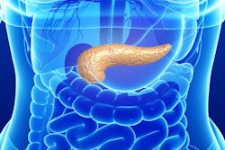
Imaging of GEP-NETs has gained growing recognition due to the clinical utilization of Ga-68-labeled somatostatin analogue PET tracers, which target the overexpressed somatostatin receptor subtype 2 (SSTR2) on the tumor cell membrane.
 Schematic representation of the mechanism of action of SSTR2 imaging. Both radionuclides -- indium-111 (In-111) and Ga-68 -- have been used for targeting imaging and therapeutics in conjunction with SSTR2 ligands. DOTA = dodecane tetra-acetic acid. DTPA = diethylenetriaminepentaacetic acid.
"While hepatic and nodal metastases are commonplace at the time of NET diagnosis, cardiac metastases are found much less frequently," explained Renfrew, who is a postgraduate year 5 (PGY-5) radiology resident. "The diagnostic criterion of cardiac metastasis is focal tracer uptake in the myocardium."
The group evaluated the prevalence, distribution pattern, and radiotracer uptake characteristics of cardiac metastasis on Ga-68 DOTATATE PET/CT. On PET images, they documented and analyzed the maximum standardized uptake value of the cardiac metastasis and left ventricular chamber (blood pool), the Krenning score of the cardiac metastasis, and the presence of multisystem metastasis. They performed correlation with diagnostic CT and/or cardiac MRI to confirm the location of metastasis in the heart.
Over 1,400 scans
Between October 2017 and March 2020, 1,426 Ga-68 DOTATATE PET/CT and PET/MRI scans were performed at the Mayo Clinic in Arizona. Twenty-six patients (mean age, 64 ± 10.17 years; male to female ratio of 11-to-10) were diagnosed with a cardiac metastasis, always with coexistence of multisystem metastases.
Of these 26 patients, three had one other organ system (bone, lymph node, pancreas, liver, bone, esophagus, orbit) involved, while nine had two other organs involved, seven had three others, five had four others, and two had five others. None of the patients exhibited relevant cardiovascular symptoms through chart review.
A total of 16 cardiac lesions were located in the left ventricle, eight were in the interventricular septum, and five were in the right ventricle.
The average SUVmax of these lesions was 9.2 (range, 2.6-24.1), and the Krenning score was 2-4. The average SUVmax of blood pool was 1.2 (range, 0.5-2.4). The average lesion to blood pool SUV ratio was 8.3 (range, 2.6-18.2).
Of the 26 cases, 17 also underwent a contemporaneous chest CT exam. Only one of these CT scans demonstrated evidence of neuroendocrine cardiac metastasis, Renfrew and colleagues reported.
 Ga-68 DOTATATE PET/CT image for a patient with a neuroendocrine metastasis in the right ventricle, ventricular apex, and left ventricular free wall.
 T2 fat-suppressed axial MRI of same patient demonstrates increased T2 signal within the cardiac metastases. The lesions also showed restricted diffusion.
 Contrast-enhanced chest CT of same patient demonstrates three filling defects corresponding to the neuroendocrine metastases seen on Ga-68 DOTATATE PET/CT and cardiac MRI. This was the only patient demonstrating a correlate on CT.
Five of the 26 patients also received a contemporaneous cardiac MRI. Four of these scans correctly identified cardiac neuroendocrine involvement.
Seven patients had multiple Ga-68 DOTATATE PET/CT scans while on octreotide therapy. Two of them showed initial progression, with subsequent lesion stabilization on imaging.
The clinical significance of cardiac metastasis needs to be further investigated, the authors stated.
"We are always increasing our number of cardiac metastases with good DOTATATE correlates," Renfrew told AuntMinnieEurope.com. "We are currently working with the cardiology department correlating our current findings with those on echocardiography."