Computer-aided detection (CAD) and risk stratification are useful for excluding lung cancer on thoracic CT, according to Scottish and Dutch researchers. CAD followed by radiology review of suspicious cases is cost-effective and can lead to a 30% reduction in screening costs, they say.
Initial screening chest CT scans from patients in Glasgow, U.K., enrolled within the prospective Early Cancer Detection test -- Lung Cancer Scotland (ECLS) study were sent for Lung-RADS analysis by the Diagnostic Image Analysis Group in the department of radiology and nuclear medicine at Radboud University Medical Center in the Netherlands.
The team used CAD to identify nodules and then categorize them into one of four groups (CAD 1-4) based upon probability of malignancy and taking into account lesion morphology, size, location, and texture. Independently and blind to the CAD results, two experienced senior respiratory radiologists reported the scans and assigned a similar grading category.
The patients were then followed up with interval CT scans, as specified in the ECLS study protocol, noted Dr. Mark Hall, a radiologist at Glasgow Royal Infirmary, and colleagues in an ECR 2018 e-poster.
CAD categorization had a statistically significant correlation with the radiologist categorization (p < 0.001), and none of the 113 (36%) patients within the CAD 1 group (no nodule identified) was found to have lung nodules, they added.
| CAD scoring compared with radiologist scoring | ||
| CAD categories | Radiologist category 1 |
Radiologist categories 1b-4a |
| CAD category 1 | 113 | 37 |
| CAD categories 2-4 | 56 | 107 |
Lung cancer is the most common cancer worldwide, accounting for 1.6 million deaths worldwide in 2012, according to the International Agency for Research on cancer, and several large U.S. institutions have recommended lung cancer screening with low-dose CT in specific population groups, the authors wrote.
"If lung cancer screening was implemented in Scotland, there would be a very large increase in requirement for chest CT scans. These would all need to be reported, increasing radiological workload and cost," they continued. "We postulated that combining the use of CAD and analysis could safely exclude lung cancer in a large number of scans, obviating the need for radiological review with associated resource savings."
Their hypothesis was that CAD would have a 100% negative predictive value for lung cancer screening.
No national scheme
Currently, the U.K. has no national screening program for lung cancer because it isn't clear that screening can save lives from lung cancer and the tests have risks and can be expensive, according to Cancer Research UK.
"Low-dose CT scans are being looked as a possible screening test for lung cancer. Tests like this have risks. The lungs are very sensitive to radiation, and frequent scans might cause lung damage," the organization noted. "Tests can also find lung changes that look like cancer and need to be checked by further tests, such as a biopsy. These further tests can also have risks."
What's more, lung screening might cause overdiagnosis, meaning that some people will go on to have lung cancer treatment that they would never have needed, and they have to cope with the side effects and anxiety that patients receiving cancer treatment go through. Researchers need to balance the benefits of a possible screening program with the risk of overdiagnosis, Cancer Research UK stated.
Expiratory radiographs: Any value?
Another thoracic study presented by Hall and his colleague Dr. John Maguire at ECR 2018 attempted to answer the question: Has the expiratory radiograph passed its expiry date?
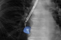
At the Glasgow Royal Infirmary, all patients with a suspected pneumothorax are investigated with both inspiratory and expiratory radiographs, but experience suggests that expiratory radiographs provide little additional benefit to the patient and ensue additional radiation dose as well as increase departmental workload and cost, he explained.
Hall performed a retrospective evaluation of all 966 pairs of inspiratory and expiratory radiographs taken over a six-month period from August 2016 to January 2017. A total of 17 pneumothoraces were detected from the 966 pairs (1.8%). All 17 pneumothoraces were visible on the inspiratory radiograph, and the average size of the pneumothorax was 3.8 cm.
"We analyzed the radiographs for pneumothorax and ascertained whether the additional expiratory radiograph provided benefit in the diagnosis of pneumothorax. In addition, we measured the average pneumothorax size," he noted.
None of the expiratory radiographs provided any additional benefit in the diagnosis or patient management. The findings of this local study are in keeping with the literature review and suggest a change of practice should be considered. Skipping expiratory radiographs will reduce workload and also provide departmental time and financial savings, Hall concluded.
Visit the ESR website to access the e-posters on the lung CAD study and the pneumothorax study presented at ECR 2018.