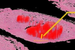
The Irish Centre for High-Performance Computing (ICHEC) has approved a third major COVID-19 research project on the national high-performance computer (HPC), known as Kay.
Led by Dr. Aaron Golden from the National University of Ireland, Galway, the team plans to develop new artificial intelligence (AI) software to expedite the diagnosis of COVID-19 from chest CT scans.
Nasal and throat swabs, the traditional methods for detecting COVID-19, are not 100% accurate, and often patients don't receive their results for a day or two. However, using a CT scan, a radiologist can tell in less than an hour if a patient has lung lesions indicative of COVID-19, Golden said.
The question becomes the following: Are the lesions due to pneumonia, a pulmonary disorder, or another lung condition? That's where AI comes in, as well as the necessity of Kay's supercomputing power. A convolutional neural network classifies CT scans into likely COVID-19 or non-COVID-19 groups, and then a deep-learning algorithm standardizes the thousands of training CT scans.
Golden's project was funded by the Health Research Board as part of the COVID-19 Pandemic Rapid Response Funding Call.




![A normal mammogram confirmed by three-year radiologic follow-up illustrates reader-marked regions of interest (ROIs) during (A) unaided (round 1) and (B) artificial intelligence (AI)–assisted (round 2) reading. Each colored dot represents an ROI for recall by a human reader. Readers could mark more than one ROI per case, represented by multiple dots of the same color. During AI-assisted reading, the AI system displayed three visible prompts: two with suspicion of malignancy scores of 35% (left mediolateral oblique [L MLO] and craniocaudal [L CC]) and one with a suspicion of malignancy score of 10% (right craniocaudal [R CC]), shown as polygonal overlays. Without AI, six of 10 readers (60%) marked a false-positive ROI. With AI assistance, this fell to two of 10 (20%). R MLO = right mediolateral oblique.](https://img.auntminnieeurope.com/mindful/smg/workspaces/default/uploads/2026/07/2026-07-14-radiology-mammogram-ai-auto-bias.H0bYO8QlWs.jpg?auto=format%2Ccompress&fit=crop&h=100&q=70&w=100)


![A normal mammogram confirmed by three-year radiologic follow-up illustrates reader-marked regions of interest (ROIs) during (A) unaided (round 1) and (B) artificial intelligence (AI)–assisted (round 2) reading. Each colored dot represents an ROI for recall by a human reader. Readers could mark more than one ROI per case, represented by multiple dots of the same color. During AI-assisted reading, the AI system displayed three visible prompts: two with suspicion of malignancy scores of 35% (left mediolateral oblique [L MLO] and craniocaudal [L CC]) and one with a suspicion of malignancy score of 10% (right craniocaudal [R CC]), shown as polygonal overlays. Without AI, six of 10 readers (60%) marked a false-positive ROI. With AI assistance, this fell to two of 10 (20%). R MLO = right mediolateral oblique.](https://img.auntminnieeurope.com/mindful/smg/workspaces/default/uploads/2026/07/2026-07-14-radiology-mammogram-ai-auto-bias.H0bYO8QlWs.jpg?auto=format%2Ccompress&fit=crop&h=112&q=70&w=112)